


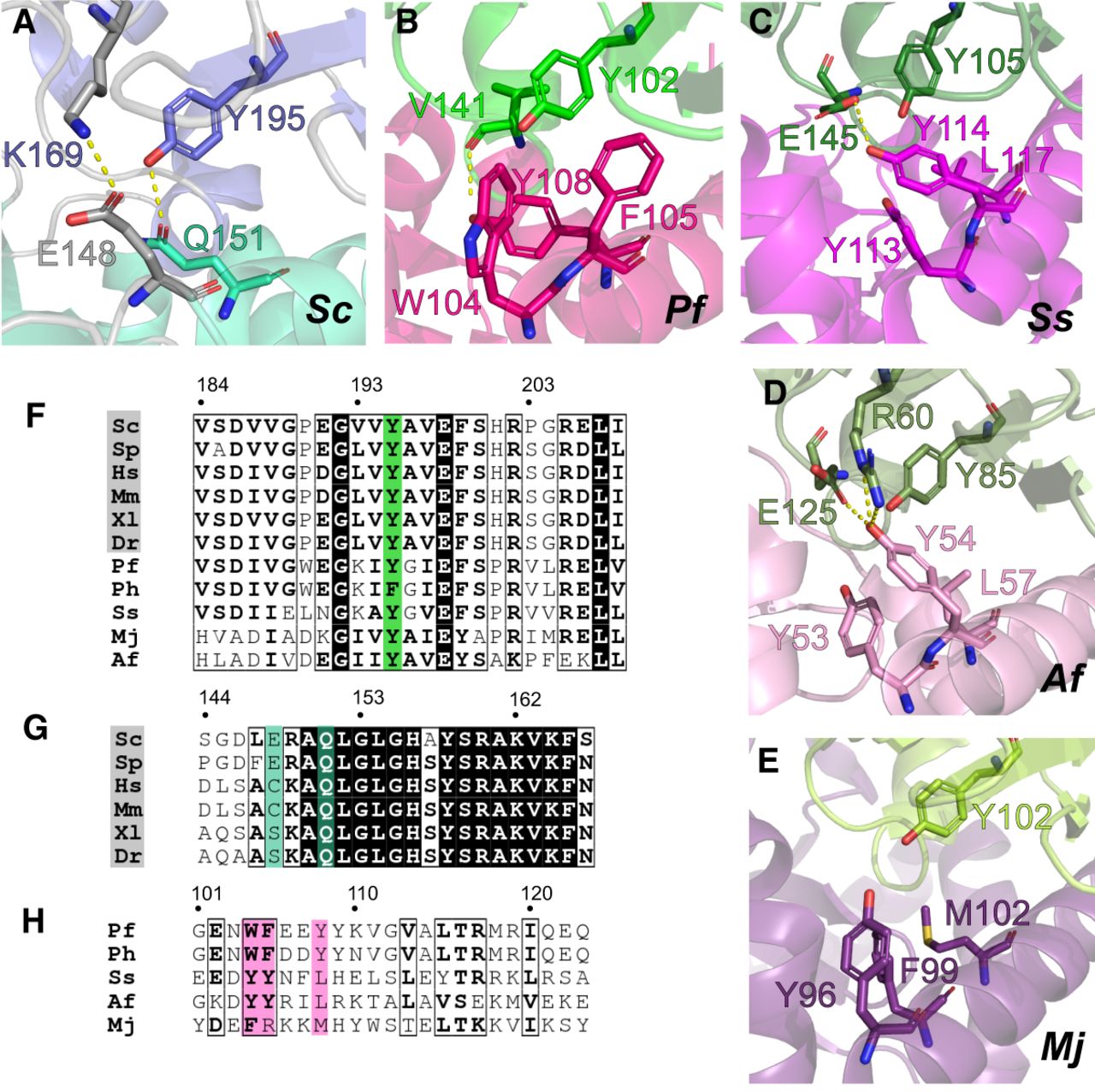
The interactions of a conserved tyrosine residue differ between archaea and eukaryotes. (A) Interactions of Nop183–327-Y195 involving residues of Nop561–166-α7. (B–E) Conserved hydrophobic clusters around the conserved tyrosine in the complexes of archaeal Fibrillarin and Nop5: (B) Pf Nop51–123–Fibrillarin (PDB-ID: 2NNW); (C) Ss Nop51–132–Fibrillarin (PDB-ID: 3ID5); (D) Af Nop51–71–Fibrillarin (PDB-ID: 1NT2); (E) docking model of the complex between the Nop5 amino-terminal domain and Fibrillarin from M. janaschii (PDB-ID: 3T7Z and 1G8S). (F) Multiple sequence alignment of the fragment from eukaryotic and archaeal Fibrillarins containing the conserved tyrosine residue (highlighted in green). Residue numbers refer to the first line of the alignment. (G) Multiple sequence alignment of residues in eukaryotic Nop56-α7 that interact with the conserved tyrosine residue in Nop1/Fibrillarin. Conserved interacting residues are highlighted in turquoise. (H) Multiple sequence alignment of residues in archaeal Nop5-α6 that interact with the conserved tyrosine residue in Fibrillarin. Conserved interacting aromatic residues are highlighted in pink.




