


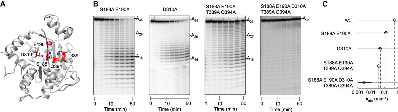
FIGURE 7.
Exonuclease activity of mutant Pop2p variants. (A) Structure of the S. cerevisiae Pop2p exonuclease domain (PDB: 1UOC). Blue spheres represent the Mg2+ ions extrapolated from the S.p. Pop2p exonuclease domain (PDB: 2P51). Amino acids in the catalytic center are marked red. (B) Representative PAGE for exonuclease reactions of Pop2p variants (as indicated) (1000 nM) with A36 (1 nM). Aliquots were removed at 1, 5, 10, 20, 30, 40, and 50 min. (C). Observed rate constants (removal of first nucleotide) for degradation of A36 with the Pop2p variants indicated. Error bar represents the standard error from fitting time courses to rate equations.




