


Cover image
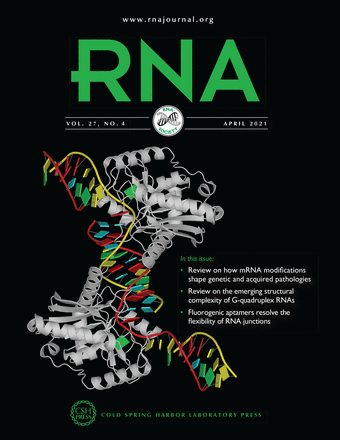
Mouse endonuclease V in complex with 23mer RNA (PDB id: 6OZO; Wu J, Samara NL, Kuraoka I, Yang W. 2019. Evolution of inosine-specific endonuclease V from bacterial DNase to eukaryotic RNase. Mol Cell 76: 44). Endonuclease V cleaves the second phosphodiester bond 3′ to a deaminated adenosine (inosine). Although highly conserved, EndoV change substrate preference from DNA in bacteria to RNA in eukaryotes. RNA backbone is displayed as red and yellow ribbons; block bases use NDB colors: A—red, C—yellow, G—green, U—cyan; protein is displayed as a white ribbon. Cover image provided by the Nucleic Acid Database (ndbserver.rutgers.edu). Image generated using DSSR and PyMOL (Lu XJ. 2020. Nucleic Acids Res 48: e74).


Molecular Cloning
The New Edition
Buy Now and Save 30%
Paperback only
(limited time offer)

|
Molecular Cloning: A Laboratory Manual (Fourth Edition) |