


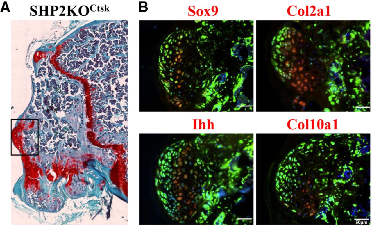
FIGURE 4.
Pathology of the tibial exostoses from SHP2KOCtsk mice. (A) Safranin O and Fast Green staining of the tibias from SHP2KOCtsk mice indicated cartilaginous exostoses. (B) RNAscope images of the magnified area (black box in A) revealed that the exostoses originated from Ctsk+ cells and expressed Sox9, Col2a1, Ihh, and Col10a1 in different regions of the exostoses. This suggested that the exostoses were benign ecchondromas. The length of the scale bar represents 50 µm.




