


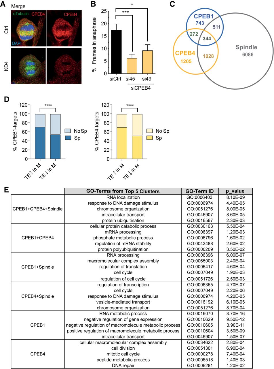
Spindle-localized mRNAs targets of CPEB1 and CPEB4 have sequential cell-cycle related functions. (A) Immunofluorescence for CPEB4, α-tubulin, and DAPI in shCtrl (Ctrl) and shCPEB4 (KD4) hTERT-RPE1 cells. (B) Quantification of percentage of frames in anaphase from total frames in mitosis for RPE1 cell transfected with a control siRNA (siCtrl) or two siRNAs against CPEB4 (SiCPEB4 #1 and #2). More than 50 cells per condition were quantified. (****) P < 0.0001, (**) P < 0.005 (Mann–Whitney test). (C) Venn diagram showing RIP targets of CPEB1 (P-value <0.05), CPEB4 (P-value <0.05), and mRNAs localized at the spindle [log2(spindle/mitosis) > 0] in Hela S3 cells synchronized with RO3306 (9 µM, 21 h) at the G2/M border and released for 45–60 min for spindle RNA-seq or CPEB4 RIP-seq. (D) Percentage of targets from CPEB1 or CPEB4 RIP-seqs localized or not at the spindle that are translationally up-regulated or down-regulated in M versus G2 according to Tanenbaum et al. (2015). (E) Top five GO categories of biological processes based on RIP-seq and spindle-seq data analyzed with the functional annotation clustering tool from DAVID bioinformatics resources.




