


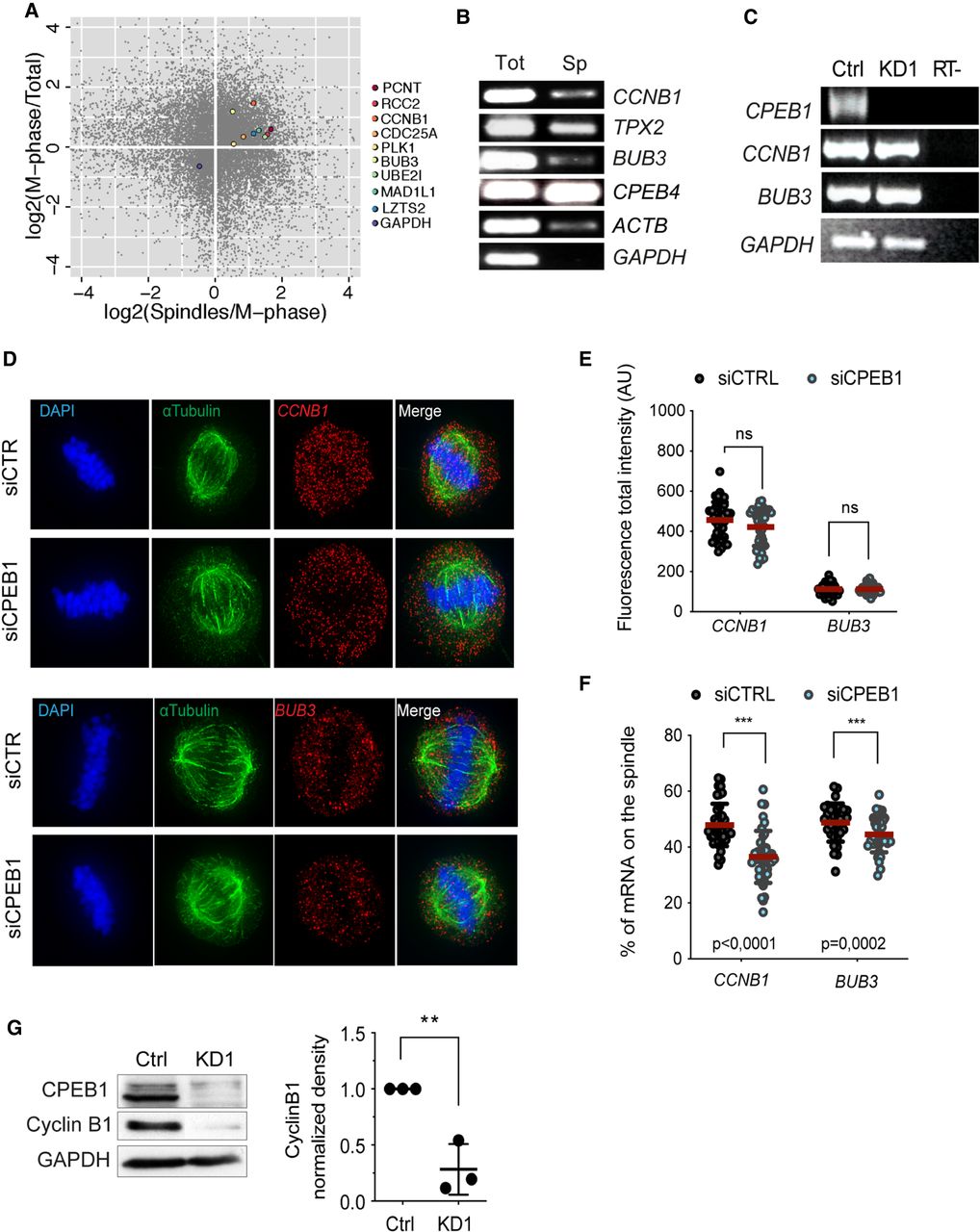
CPEB1 regulates localization at the mitotic spindle of CPE-containing mRNAs. (A) Genome-wide analysis of spindle-purified mRNAs from HeLa S3 cells, with the scatter plot showing the mRNA enrichment in M phase compared to asynchronous cells (y axis), and in spindle-associated transcripts compared to M-phase total transcriptome. (See Supplemental Table 1 for the full list of transcripts). (B) RT-PCR of the indicated mRNAs extracted from HeLa S3 cells arrested in P/M (Tot) or from purified mitotic spindles (Sp). GAPDH was used as a negative control. (C) RT-PCR of CPEB1, CCNB1, BUB3, and GAPDH mRNAs in hTERT-RPE1 control (Ctrl) or CPEB1 sh-RNA (KD1) cells. RT– indicates the control without retrotranscription. (D) smFISH images of U2OS cells treated with siRNA control (siCTRL) or siRNA CPEB1 (siCPEB1). Chromosomes and spindles were visualized by DAPI staining and α-tubulin immunofluorescence, respectively. (E,F) smFISH quantification of U2OS cells treated with siCTRL or siCPEB1. Total fluorescence intensity (E) and percentage of the indicated mRNAs localized in the spindles (F) in WT and KD1 cells is shown (see Supplemental Fig. S2C for quantification details). (G) Representative CPEB1 and Cyclin B1 western blot for WT and KD1 hTERT-RPE1 cells (left) and quantification of three independent experiments (right).




