


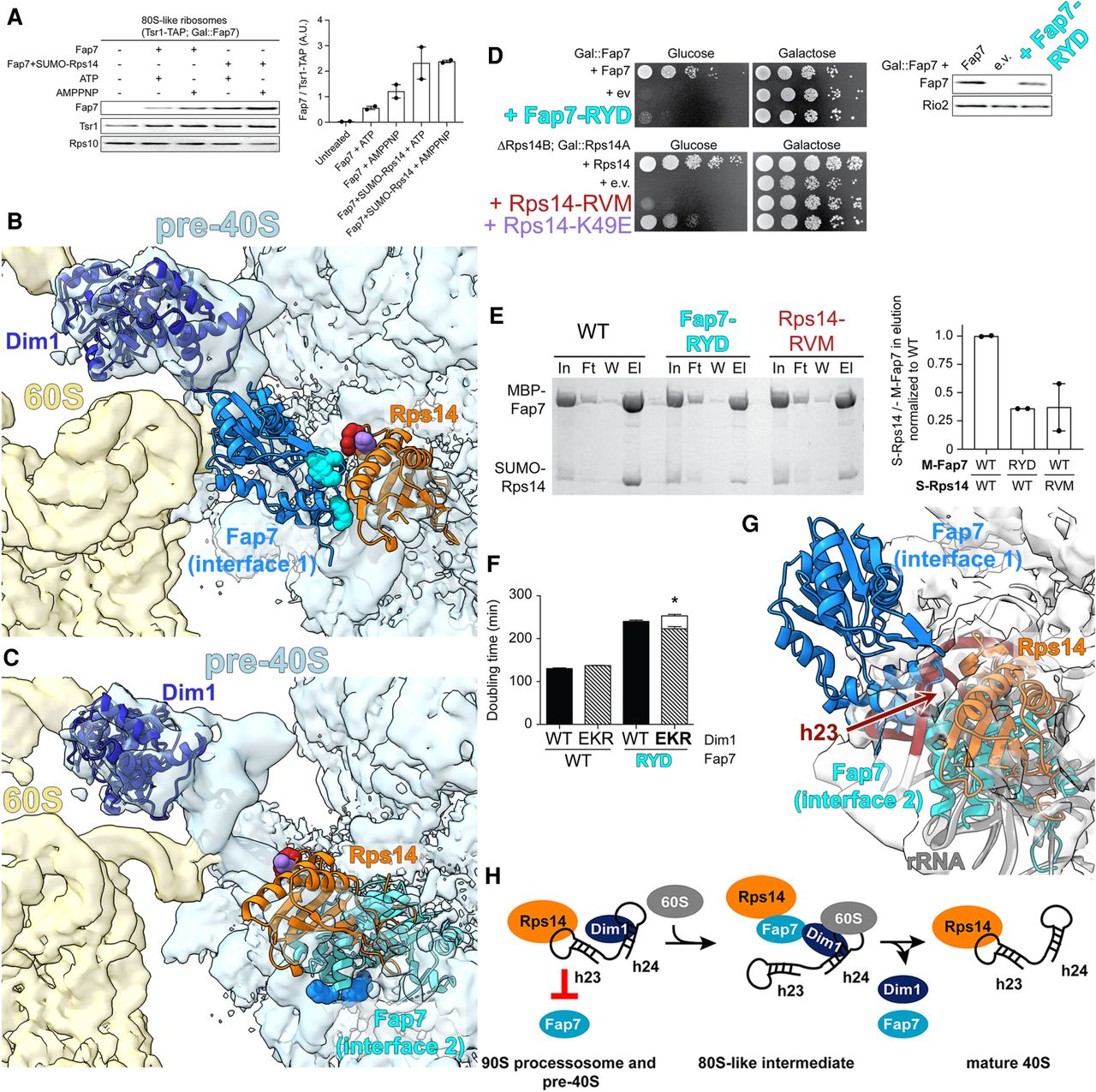
Fap7 can only bind an opened platform. (A) Fap7 binds to purified 80S-like ribosomes. Fap7 binding in the presence and absence of Rps14 and ATP or AMPPNP was assessed in a pelleting assay. Ribosome pellets were probed for Fap7, Tsr1-TAP or Rps10 (left), and bound Fap7 was quantified relative to the amount of 80S-like ribosomes (Tsr1-TAP, right). Data are the average of two replicates and error bars indicate SEM. Arbitrary Units, A. U. (B) Docking Fap7•Rps14 (Fap7, chain G and Rps14, chain F) from PDB ID 4CW7 (Loc'h et al. 2014) onto Rps14 (orange) in 80S-like ribosomes places Fap7 (bright blue) in direct contact with Dim1 (dark blue), as predicted by previous biochemical data (Ghalei et al. 2017). The residues in Fap7-RYD (R114E/Y116A/D118K, cyan) and Rps14-RVM (R41E/V42L/M46A, red) are at the interface between Fap7 and Rps14 (formed between chains G and F). Rps14-K49E is in purple. In contrast, chain C from that same structure clashes with the pre-40S platform. (C) An alternative dimer of Fap7•Rps14 reveals that Fap7 (cyan) would be in steric clash with the pre-40S platform. (D) Growth (left) or Fap7 expression levels (western blot, right) of Gal::Fap7 cells containing an empty vector (e.v.), wild-type Fap7, or Fap7-RYD or growth of ΔRps14B; Gal::Rps14A cells containing an empty vector (e.v.), wild-type Rps14, Rps14-RVM, or Rps14-K49E plasmids were compared by 10-fold serial dilution on YPD or YPGal plates. (E) Interface mutations in Fap7 or Rps14 weaken their binding affinity for each other. Shown are Coomassie-stained SDS-PAGE gels of protein binding assays on amylose beads of purified, recombinant MBP-Fap7 or MBP-Fap7-RYD and SUMO-Rps14 or SUMO-Rps14-RVM. In, input; Ft, flow-through; W, final wash; El, elution (left). Quantification of SUMO-Rps14 (S-Rps14) compared to MBP-Fap7 (M-Fap7) in elution normalized to wild-type (right). Data are the average of two replicates and error bars indicate SEM. (F) Doubling time, in minutes, of cells depleted of endogenous Dim1 and Fap7 (Gal::Dim1; Gal::Fap7) and transformed with plasmids encoding Dim1 or Dim1-EKR and either Fap7 or Fap7-RYD. The white column represents the expected doubling time if there was no rescue of Fap7-RYD by Dim1-EKR (See also Ghalei et al. 2017). The height of this column was calculated by multiplying the observed fold differences for each single mutation. The data are the average of 16–17 biological replicates and the error bars represent SEM. Unpaired t-test was performed comparing expected and actual doubling times of cells expressing Fap7-RYD and Dim1-EKR. (*) P-value = 0.030. (G) Both Fap7•Rps14 interfaces clash in pre-40S ribosomes (Rps14 [orange], h23 [red], rRNA [gray] from PDB 6FAI/EMD-4128 [Scaiola et al. 2018]). (H) The Fap7 binding site on Rps14 is blocked by h23 in nucleolar 90S preribosomes and pre-40S ribosomes.




