


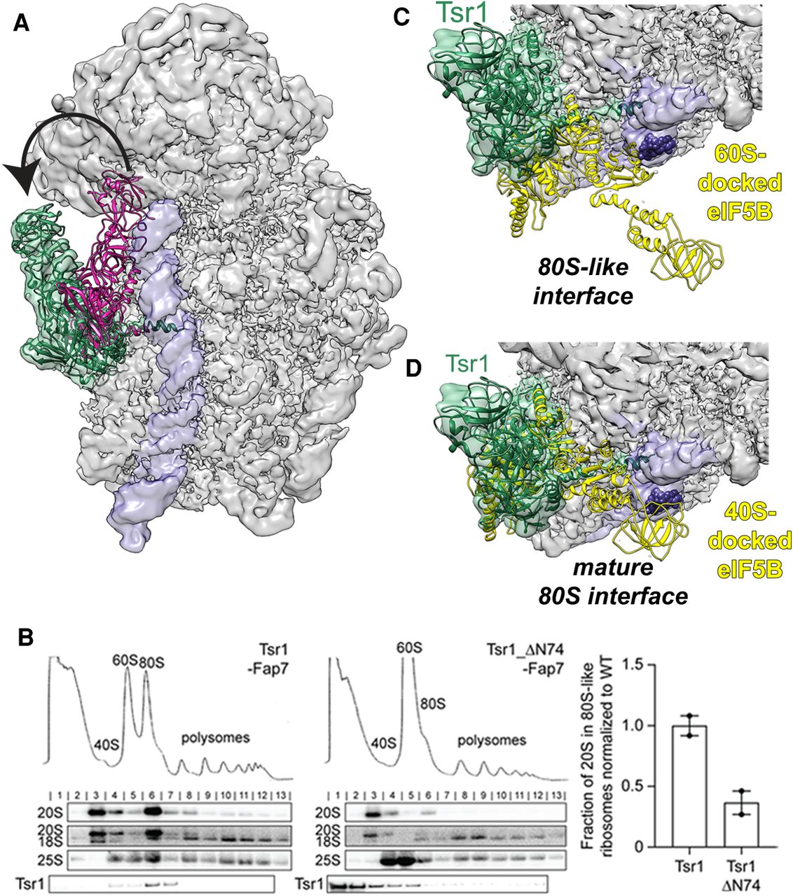
Tsr1 is repositioned to the beak. (A) The position of Tsr1 in 80S-like ribosomes (green) differs from the position in an earlier cytoplasmic pre-40S intermediate (pink, from EMD-8349 [Johnson et al. 2017]). h44 is shown in purple. (B) Whole cell extracts were fractionated as in Figure 3 (left). Quantification of the gradient northern blots (right). Fraction of 20S in 80S-like ribosomes (fractions 6–7) compared with total 20S was calculated. Data are the average of two biological replicates, normalized to wild-type Tsr1, and error bars indicate the SEM. (C) The opened pre-40S and 60S interface leaves space for eIF5B (yellow) across h44 (purple), blocking the early-forming B3 bridge (marked by nucleotides 1655–1657, shown as purple spheres). Model was obtained by superimposition of the 60S subunits from 80S-like ribosomes and the eIF5B-bound mature 80S ribosome (PDB ID 4V8Z [Fernández et al. 2013]). (D) If subunits were joining in the canonical mature 80S-structure, Tsr1 binding would block eIF5B recruitment. Model was obtained by superimposition of the 40S subunits from 80S-like preribosomes and the eIF5B-bound mature 80S ribosome (PDB ID 4V8Z [Fernández et al. 2013]). The clash score, defined in Phenix (Adams et al. 2010) as the number of overlaps greater than 0.4 Å/1000 atoms, increases from 170 (60S superimposition) to 780 (40S superimposition), an increase from 2% to 11% of the atoms in Tsr1.




