


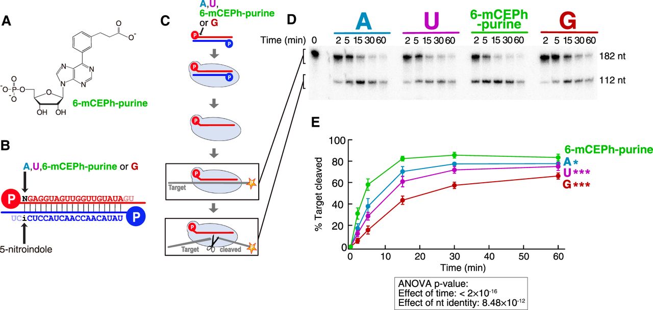
The 6-mCEPh-purine modification improves the target cleavage. (A) Chemical structure of the 6-mCEPh-purine modification. (B) Four siRNA duplexes used in this set of experiments. They bore different nucleotides (A, U, 6-mCEPh-purine or G) at the 5′ end of the guide strand and 5-nitroindole at position 19 of the passenger strand. Both the guide and passenger strands had a 5′ monophosphate. (C) Scheme of RISC assembly and target cleavage. The target RNA was 5′ cap-radiolabeled. (D) A representative result of the target cleavage assay. The upper and lower bands correspond to the full-length and cleaved target, respectively. (E) Quantification of the target cleavage assay. The graph shows the average ± SD from three independent experiments using the same set of reagents. The ANOVA P-values are given in an inset under the graphs. The results of pairwise comparison on the effect of 6-mCEPh-purine compared to natural nucleotides are indicated by asterisks (*) P < 0.05, (**) P < 0.005, (***) P < 0.0005. The detailed results of statistical analyses are summarized in Supplemental Table 1.




