


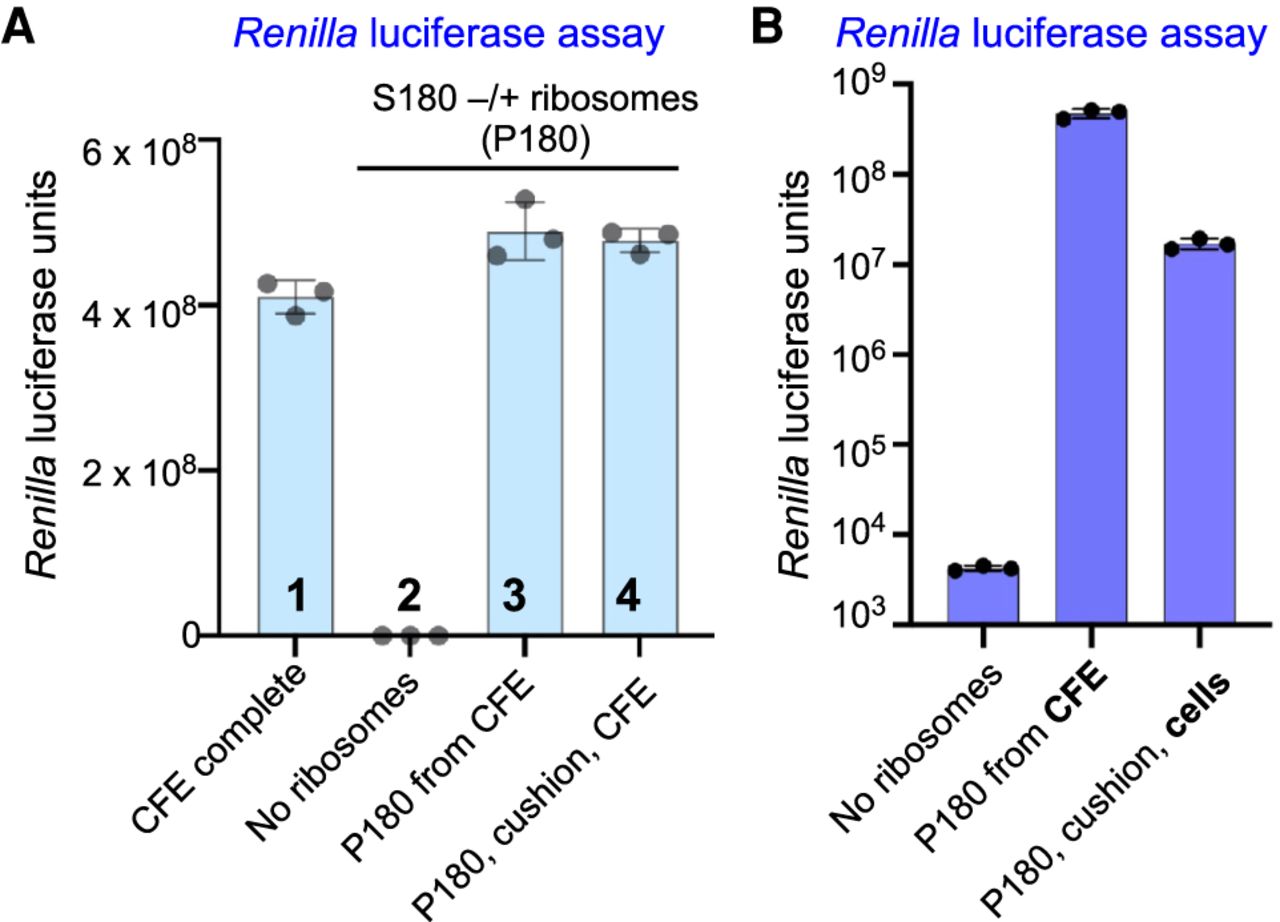
(A) Centrifugation of ribosomes through glycerol cushion does not affect their translational activity. Translation reactions were assembled with complete CFE (lane 1) or with 9 µg of purified ribosomes in the RSR format (lanes 3–4). For RSR, ribosomes were pelleted directly from CFE (lane 3) or by centrifugation of CFE through a 20% glycerol cushion (lane 4). In lane 2, no ribosomes were added to the reaction. (B) Activity of ribosomes prepared from CFE and from cells lysed by glass-bead beating. RSR translation reactions were assembled with 9 µg of ribosomes pelleted from the complete CFE (P180 from CFE) or with 9 µg of ribosomes purified from cell lysate by 180K-centrifugation through 20% glycerol cushion (P180, cushion, cells). In the control reaction, no ribosomes were added, and the background levels of luminescence are detected. In A and B, each reaction contained 400 ng of capped TAP-RLuc mRNA reporter. Reactions were incubated at 21°C for 90 min and the reaction products were analyzed by a Renilla luciferase assay. Error bars represent SEM of three experiments.




