


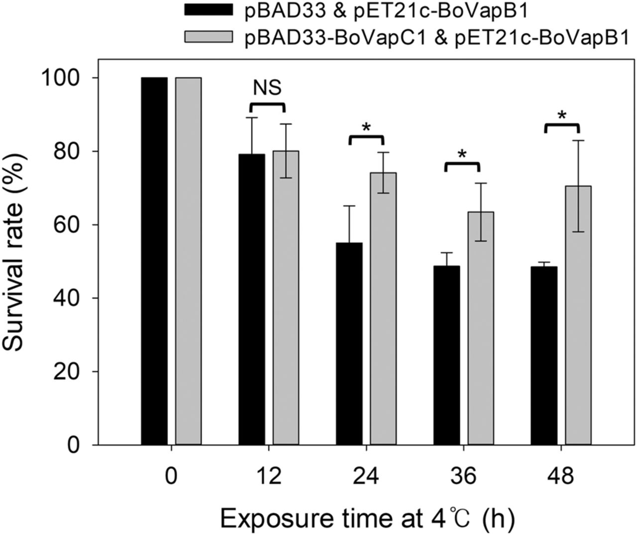
FIGURE 7.
Survival rates of cells expressing BoVapC1 after exposure to 4°C. The survival rates were obtained by calculating the percentage of the number of CFU in the BoVapC1-induced cells, compared with those in control cells. Experiments were performed in three independent repetitions and error bars represent SD. Statistical significance was derived from the two-tailed t-test. NS, nonsignificant; (*) P < 0.05.




