


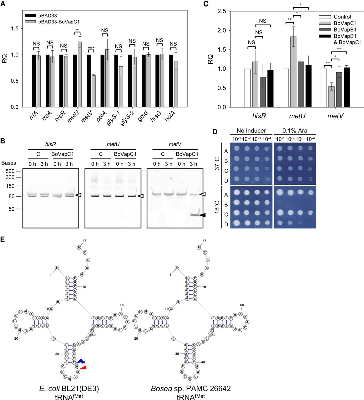
tRNAfMet-specific cleavage activity of BoVapC1. (A) qRT-PCR analyses of the level of mRNAs, rRNAs, and tRNAs. BL21(DE3) cells harboring pBAD33 or pBAD33-BoVapC1 were cultivated as described in Materials and Methods. Total RNAs extracted from these cells were utilized for qRT-PCR analyses. Experiments were independently conducted three times, and error bars represent SD. NS, nonsignificant; (*) P < 0.05; (***) P < 0.001. (B) Northern blotting analyses of tRNAs. RNAs were extracted from BL21(DE3) cells carrying pBAD33 (C) or pBAD33-BoVapC1 (BoVapC1) before (0 h) and after induction (3 h) and blotted with biotin-labeled oligonucleotides which are specific to the tRNAs as described in Materials and Methods. Full-length or cleaved tRNAs are indicated with open or filled arrowheads, respectively. Low Range ssRNA Ladder (New England Biolabs) was used as a size marker. (C) Inhibition of BoVapC1 ribonuclease activity by BoVapB1. BL21(DE3) cells harboring plasmid pairs were cultivated as described in Materials and Methods. Total RNAs extracted from these cells were utilized for qRT-PCR analyses. Experiments were independently performed three times. Error bars represent SD. NS, nonsignificant; (*) P < 0.05; (**) P < 0.01. (D) Growth restoration by overexpression of metZWV in BoVapC1-expressing cell. The BL21(DE3) cells pretransformed with pBAD33 or pBAD33-BoVapC1 were double transformed with pBR322 or pBR322-metZWV. The spotting assay was conducted as described in Figure 2. A; cells with pBAD33/pBR322, B; pBAD33-BoVapC1/pBR322, C; pBAD33/pBR322-metZWV, and D; pBAD33-BoVapC1/pBR322-metZWV. (E) The sequences and secondary structures of tRNAfMets. The structures of tRNAfMet of E. coli BL21(DE3) and Bosea sp. PAMC 26642 were drawn using StructureEditor (Reuter and Mathews 2010). Blue and red arrows indicate the positions cleaved by S. flexneri VapC (Winther and Gerdes 2011) and NTHi VapC (Walling and Butler 2018), respectively.




