


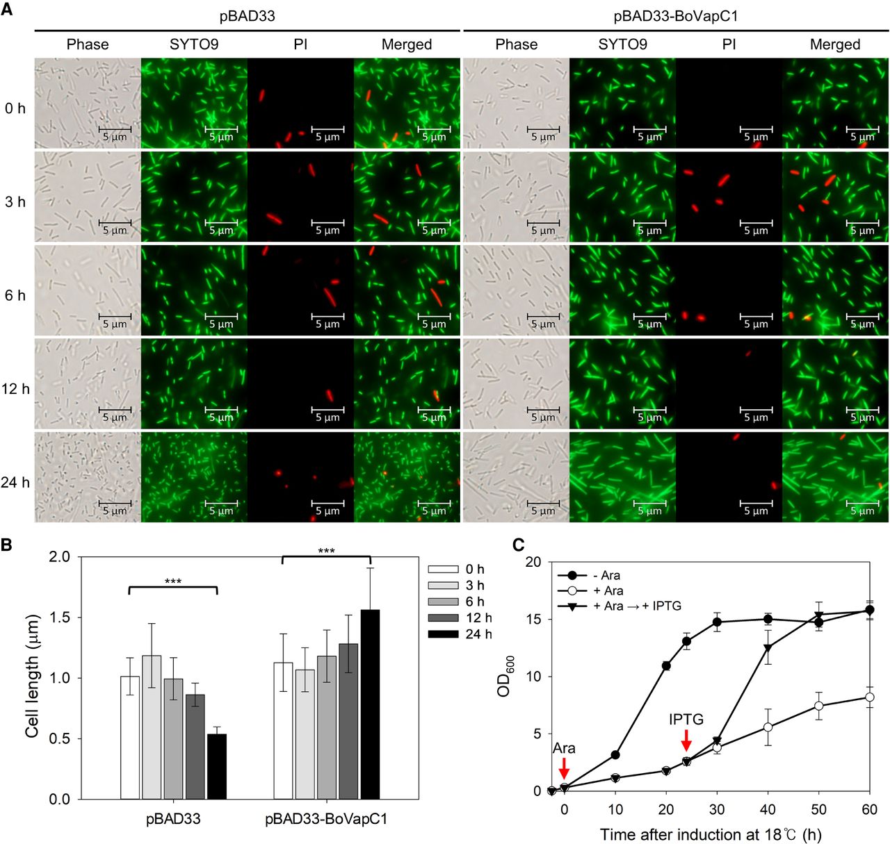
Bacteriostatic effect of BoVapC1 on E. coli growth. (A) Live/dead staining of BL21(DE3) cells expressing BoVapC1. E. coli BL21(DE3) transformants harboring pBAD33 or pBAD33-BoVapC1 were grown and stained for fluorescence microscopy as described in Materials and Methods. (B) Effect of BoVapC1 expression on the cell length of E. coli. Cell length data from Figure 5A was obtained using the MicrobeJ plugin for ImageJ (Ducret et al. 2016). One hundred and thirty seven cells were counted for each condition. Error bars represent SD. Statistical significance was derived using the two-tailed t-test. (***) P < 0.001. (C) Resumption of cell growth by antitoxin expression. BL21(DE3) cells harboring both pET21c-BoVapB1 and pBAD33-BoVapC1 were grown in M9 media containing Amp and Cm at 37°C to the early exponential phase. Then, the bacterial culture was diluted fivefold in fresh M9 media supplemented without (●) or with 0.4% arabinose, respectively, and further incubated at 18°C for 60 h (○). At 24 h after toxin induction, IPTG was added at a final concentration of 0.1 mM (▾). Red arrows indicate induction starting points. Three independent experiments were carried out, and error bars represent SD.




