


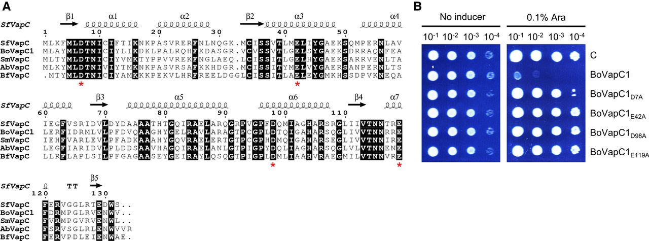
Multiple sequence alignment of BoVapC1 with previously characterized VapC homologs in other bacteria. (A) The primary sequence of BoVapC1 (AXW83_01405) was aligned with various VapC homologs in S. flexneri (Sf), S. meliloti (Sm), A. bacterium (Ab), and B. fragilis (Bf). The secondary structure of SfVapC is presented above the alignment. The acidic amino acid residues which are conserved in the active site of VapC are shown by red asterisks at the bottom of the alignment. The numbers correspond to the residues. The GenBank accession numbers for the VapC homologs are as follows: SfVapC; WP_000911329.1, SmVapC; WP_003527486.1, AbVapC; NDA60252.1, BfVapC; KAA4851476.1. (B) Effect of mutations in conserved acidic residues of BoVapC on growth-inhibitory activity. E. coli BL21(DE3) cells were transformed with pBAD33, pBAD33-BoVapC1, or pBAD33-BoVapC1 mutants. The transformant colonies were scraped and diluted as described in Figure 2. Bacterial suspensions were spotted on M9 agar plates supplemented with 0% and 0.1% arabinose. The plates were incubated at 18°C. C indicates pBAD33.




