


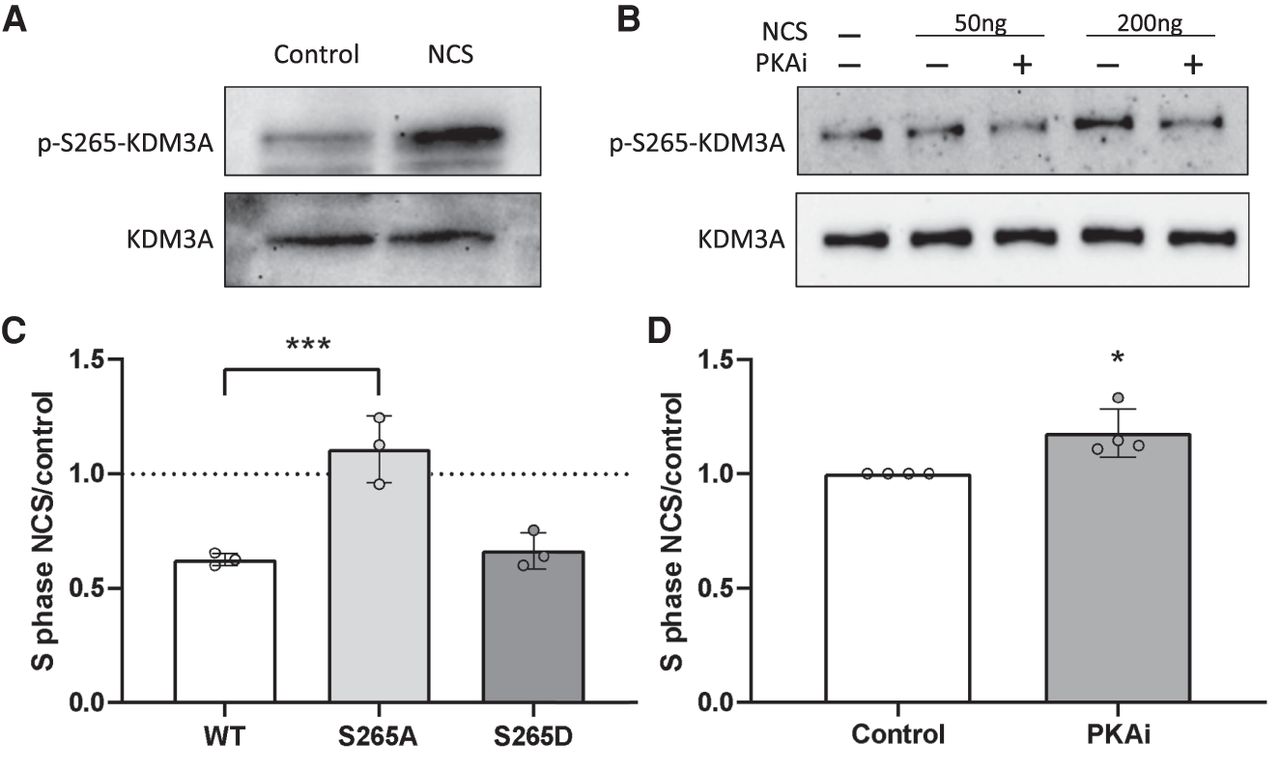
KDM3A is phosphorylated by PKA following double strand breaks. (A) MCF7 cells treated with 200 ng/mL NCS for 1 h. Immunoblotting was conducted with the indicated antibodies. (B) MCF7 cells were treated with PKAi for 30 min following treatment with 50 and 200 ng/mL NCS for 1 h. Immunoblotting was conducted with the indicated antibodies. (C) MCF7 cells stably expressing either wild-type KDM3A or KDM3A mutated at serine 265 to alanine (S265A) or to aspartic acid (S265D) were incubated with 200 ng/mL NCS, and analyzed 8 h later using flow cytometry. The amount of cells accumulated in S-phase is shown as a fold-change relative to untreated cells. (D) MCF7 cells were incubated with PKAi for 30 min followed by treatment with 200 ng/mL NCS, and analyzed 8 h later using flow cytometry. The amount of cells accumulated in S-phase is shown as a fold-change relative to untreated cells. (C,D) Plot represents the mean of four independent experiments and ± SD. (*) P < 0.05; (**) P < 0.01.




