


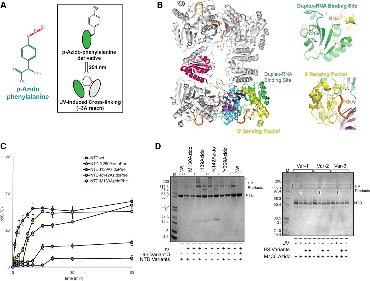
Incorporation of azido-phenylalanine into the RNase E catalytic domain. (A) Chemical formula of para-azido-phenylalanine (p-AzidoPhe); inset shows p-AzidoPhe photo-crosslinking to nearby residues upon exposure to UV light at 254 nm. (B) Models of RNase E NTD tetramer with bound RNA at active site, 5′ sensor and the duplex recognition region with insets showing the residues (M130, I139, R142, and Y269) substituted with p-AzidoPhe (model based on PDB 2C0B). (C) Time course assay of p5S production from 9S RNA, processed by p-AzidoPhe derivatives of NTD; values represent mean (n = 3) and standard deviation. (D) Denaturing protein gels showing p-AzidoPhe derivatives of RNase E NTD form UV cross-linked product(s). The p-AzidoPhe modified protein may form intradomain interaction(s) upon light exposure which are lost in the presence of 9S RNA, suggesting masking of the crosslinking moiety upon RNA binding.




