


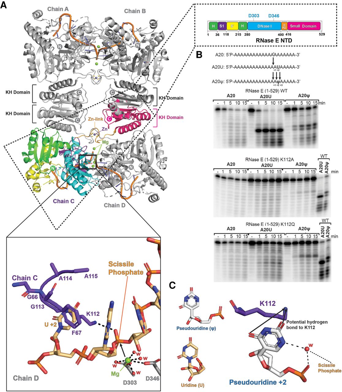
Role of RNase E K112 in interaction with uracil +2 of the substrate, and impact of pseudouridylation. (A) The tetrameric RNase E catalytic domain (NTD) in complex with RNA (PDB: 2C0B) (Callaghan et al. 2005). The inset on the upper right shows a cartoon schematic of the domains showing active-site residues D303 and D346. The lower inset shows a model of the organization of binding of RNA substrate based on the structure of chemically protected RNA (PDB 2C0B). The residues in purple are from the S1 domain of RNase E and the scissile phosphate from the RNA bound in the active site on the interface of two protomers presented for the hydrolytic attack by the waters associated (W, red) with magnesium ion (Mg++, green sphere); the U+2 is proposed to be sandwiched between side chains of amino acids K112 and F67. (B) Cleavage assays of RNase E. Cleavage of 20-mer polyadenine (A20), polyadenine with an uracil at position 15 (A20U), and polyadenine with a pseudouridine (ψ) at position 15 (A20ψ) by wild-type RNase E NTD (top panel), RNase E NTD with a mutation of K112A (middle panel), and K112Q (bottom panel). The substrate was 5′ end-labeled and the products were resolved on denaturing urea-PAGE gels. The time points of the reactions are annotated above the gels. (C) A proposed model of the likely hydration organization at the site of pseudouridine (ψ). The model also shows a probable hydrogen-bond mediated interaction between K112 and pseudouridine. A crystal structure of a duplex RNA (PDB 3CGS) was used to make the model.




