


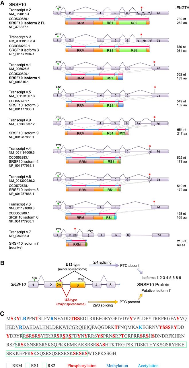
Structure of the human SRSF10 gene, transcripts and SRSF10 proteins. (A) The structure of SRSF10 mRNA variants produced by alternative splicing and corresponding protein isoform structures are shown. The length in nucleotides (nt) of the mRNA and the coding portion in amino acids (aa) are indicated, as well as their RefSeq accession numbers. Stop codons are indicated by asterisks (*). Exon 6 can be alternatively spliced to produce two forms; the shorter one lacking a serine in the RS1 domain (o). (B) Structure of the poison-exon region of SRSF10. The premature termination codon (PTC shown as *) is in the exon 2a region. (C) Known post-translational modifications on SRSF10 based on data compiled in https://www.phosphosite.org/proteinAction?id=6181&showAllSites=true.




