


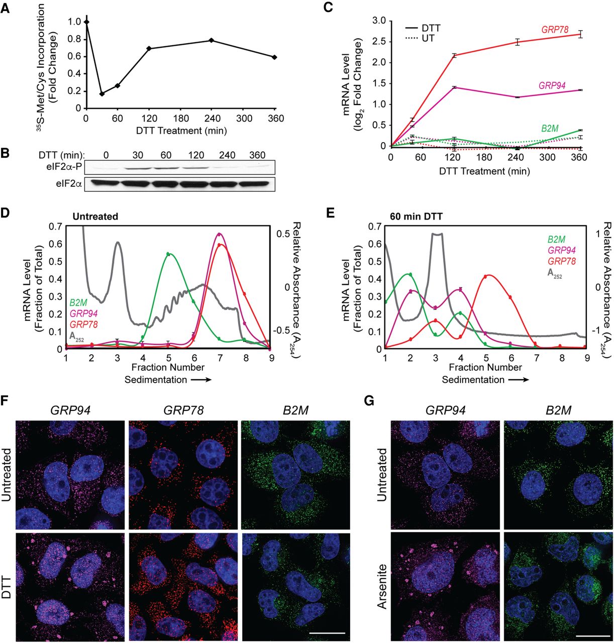
Selective recruitment of ER-targeted mRNAs into UPR-induced stress granules. (A) Representative time course of UPR-induced inhibition of protein synthesis assayed by [35S]Met/Cys incorporation. HeLa cell cultures were treated with DTT and protein synthesis rates assayed at the the indicated time points. (B) Immunoblot analysis of eIF2α and phospho-eIF2α levels following DTT treatment of cell cultures for the indicated times. (C) Representative time course of UPR-elicited transcriptional activation of the UPR response genes GRP94 and GRP78 and the ER-targeted gene B2M. Cell cultures were treated with DTT and total RNA was extracted for RT-qPCR analysis of transcript levels at the indicated time points. Data points are mean log2 fold-change ± SD, normalized to GAPDH levels. (D,E) Polyribosome profiling of GRP94, GRP78, and B2M translational status by sucrose density gradient velocity sedimentation. HeLa cell cultures at time zero (D) or following DTT treatment (E) were detergent extracted, and total polyribosome profiles were determined by the A254 nm absorbance traces (gray). mRNA distributions were determined by RT-qPCR analysis of GRP78 (red), GRP94 (magenta), and B2M (green) mRNAs extracted from the gradient fractions. Data are representative of three biological replicates; RT-qPCR data are mean fraction of total mRNA for the given gene across all gradient fractions ± SD. (F) Representative smFISH visualization of GRP94 (magenta), GRP78 (red), and B2M (green) mRNAs in untreated and DTT-treated (60 min) HeLa cells. (G) As in F but treatment with sodium arsenite (60 min). DAPI nuclear stain (blue) is indicated for all images. Scale bar = 20 µm.




