


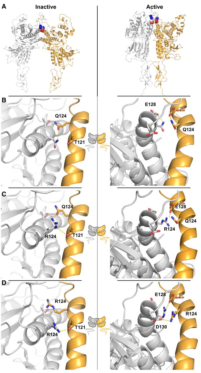
FIGURE 7.
Structural modeling of the Q124R edit site in an mGlu4 dimer. (A) mGlu4 homology model of the extracellular domains based on inactive and active mGlu5 structures (PDB: 6N50 and 6N4X). The positions of Q124 (red) and K129 (blue) are shown in spheres. Ribbon representation of Helix B and B′ (gold helix, all other secondary structure removed for clarity) of the opposite protomer in dimers with two nonedited protomers (B), a nonedited and an edited protomer (C), or two Q124R edited mGlu4 protomers (D). Dashed yellow lines show potential H-bonding interactions between side chains.




