


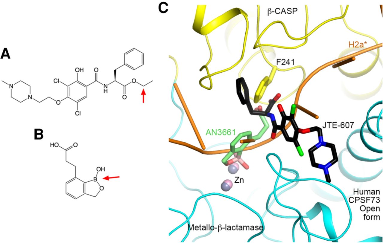
Inhibitors of CPSF73. (A) Chemical structure of JTE-607. The ester group that is hydrolyzed to produce the active acid analog is indicated with the red arrow. (B) Chemical structure of AN3661. The oxaborole moiety is indicated with the red arrow. (C) The binding modes of JTE-607 acid analog and AN3661 clash with the active, open form of human CPSF73. The structure of JTE-607 in complex with the closed form of human CPSF73 (Ross et al. 2020) is superimposed with that of the open form (Sun et al. 2020b), using the metallo-β-lactamase domain as the reference. Similarly, the structure of AN3661 in complex with the closed form of C. hominis CPSF73 (Swale et al. 2019) is superimposed with that of human CPSF73 open form. Only the inhibitors from the two structures are shown.




