


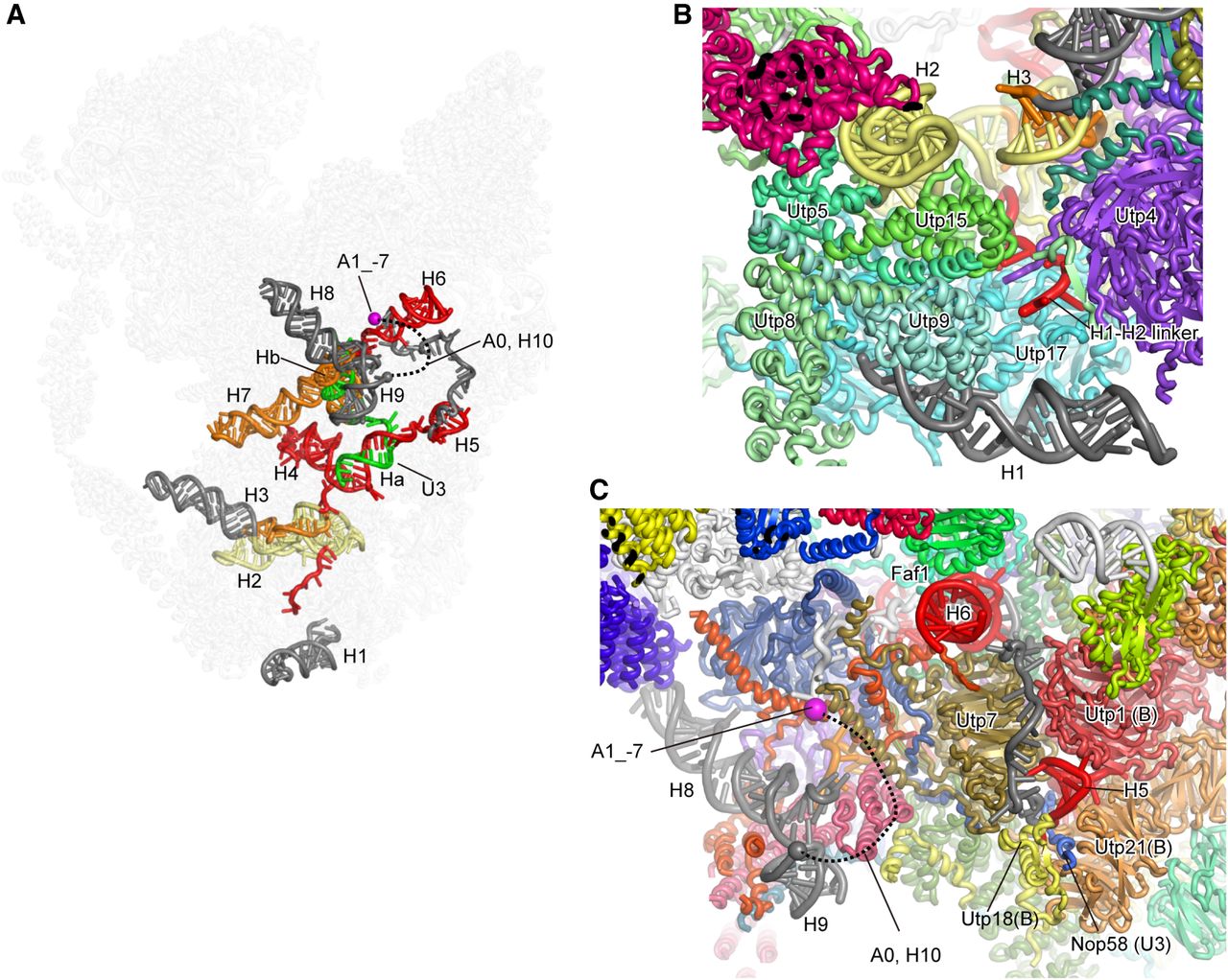
5′ ETS structure in 90S preribosome. (A) Structure of 5′ ETS in 90S. The most to least important regions are colored red, orange, yellow, and gray. The Cα atom of residue at position −7 to the A1 site is displayed as a magenta sphere. U3 snoRNA is colored in green. The A0 site and H10 are not modeled in 90S structure and shown as a dotted line connecting H9 and the A1_−7 site. The other components of 90S are shown as semitransparent background. The model is combined from two 90S structures (PDB codes 5WLC and 5WYJ). (B) 90S structure illustrating the interaction between H1–H3 and UTPA proteins. (C) 90S structure illustrating the assembly of H5, H6, H8, and H9. The unmodeled A0 site and H10 are shown as a dotted line. UTPB proteins are marked as B.




