


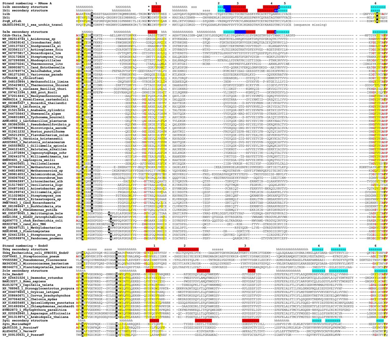
Multiple alignment of RNase A-like and EndoU-like protein sequences. Unique identifiers in the NR or PDB databases are shown before each sequence. In the secondary structure lines, h stands for a helical structure and s stands for an elongated structure (a strand). Vertebrate and sea urchin RNase A sequences are superimposed on the alignment to maximize the overlap of the structurally equivalent secondary structure elements. Conserved catalytic histidines are shown in white-on-black type, conserved hydrophobic residues (I, F, L, M, V, W, Y) are indicated by yellow shading, conserved residues with the propensity to make turns or kinks in the main chain (A, G, S, P) are indicated by bold red type, and catalytic lysine in animal RNases A is shown by asterisks in the secondary structure line. The strands that belong to the same β-sheet in EndoU family, or to the same wing of a sheet in RNases A, are shaded with the same color, and dark blue shading indicates the crease connecting the two wings in RNases A. The numbering of the β-strands is modified to consider only structurally equivalent strands, accounting for the contributions of the long strand to both wings in RNases A (see text for details).




