


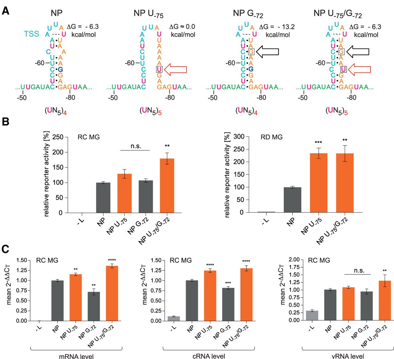
UN5 hexamer continuity between PE1 and PE2 increases viral transcription and replication. (A) Sequence and predicted structure of the wt NP HP (left), variant NP U−75 and the stabilized variants NP G−72 and NP U−75/G−72; the red arrow indicates the mutation that extends UN5 hexamer phasing and the black one marks the mutation at position −72 that stabilizes the HP stem by converting the A,C mismatch to a G:C bp. The number of UN5 hexamers between nt −51 and −80 is indicated below each individual structure. The ΔG values of the stem–loops are the minimum free energies of the centroid secondary structures predicted by RNAfold for the sequence of nt −52 to −81 using the default parameters (http://rna.tbi.univie.ac.at/cgi-bin/RNAWebSuite/RNAfold.cgi). The dashed line connecting A−65 and U−69 indicates base-pairing as suggested by RNAfold. (B) Corresponding luciferase activities of RC (left panel) or RD (right panel) MGs carrying the NP variants illustrated in panel A. Activity values are given in % relative to the native 3′-leader (NP [wt] = 100%). As a negative control, the plasmid encoding the L gene was omitted (−L) during transfection. (C) Corresponding levels of viral mRNA, cRNA, and vRNA measured by a two-step strand-specific qRT-PCR of RC MG samples using the same cells as in panel B. For more details, see Materials and Methods. In panels B and C, mean 2−ΔΔCT values (±SEM) were derived from three independent experiments with at least three technical replicates each. (**) P < 0.01; (***) P < 0.001; (****) P < 0.0001; n.s., not significant (unpaired t-test).




