


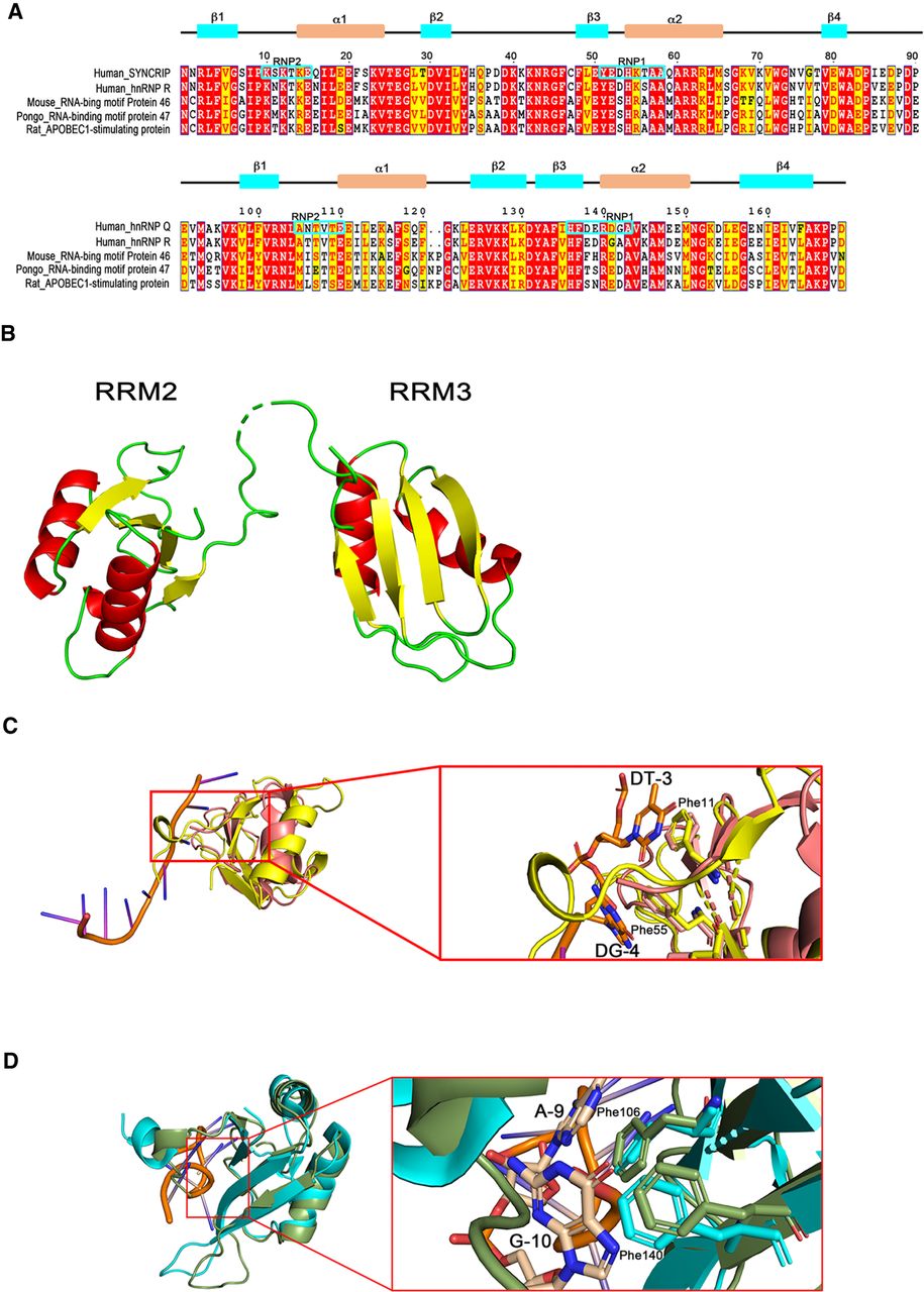
FIGURE 6.
The structure of RRM2–3 from SYNCRIP. (A) Sequence alignment of RRM2–3 domain with various RNA binding domains from RBPs. The upper panel shows the secondary structure of RRM2–3. (B) Crystal structure of RRM2–3 at 2.5 Å resolution, showing characteristic RRM folding as β1α1β2β3α2β4 topology. (C) Structure alignment of SYNCRIP RRM2 domain (yellow) with mouse TDP-43 RRM2 domain (salmon; PDB entry 3D2W). (D) Structure alignment of SYNCRIP RRM3 (green) with human hnRNP A1 RRM 2 (cyan; PDB entry 6DCL).




