


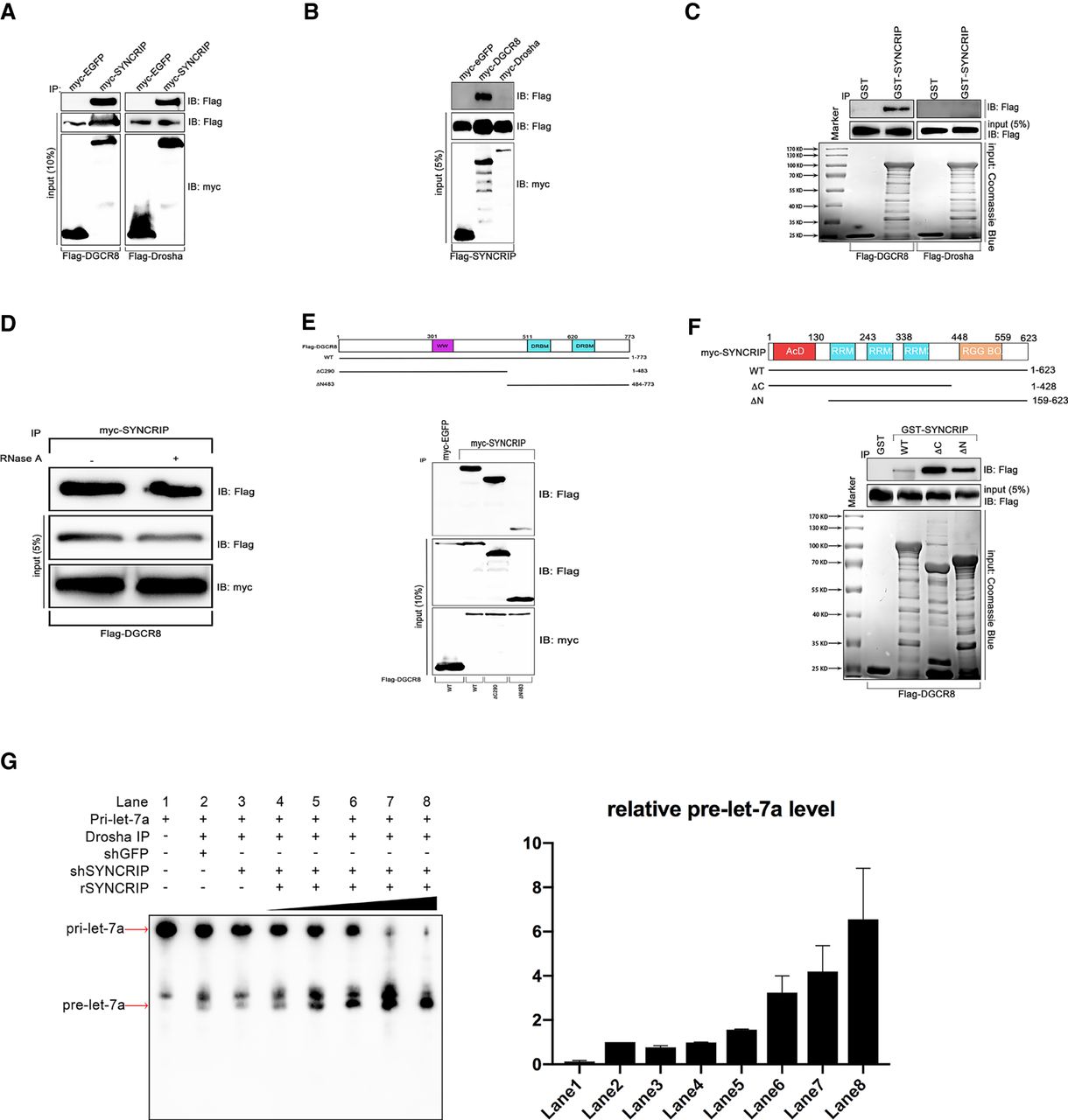
SYNCRIP associates with microprocessor complex and promotes pri-let-7a processing. (A) Identification of the interaction of SYNCRIP with DGCR8 and Drosha through Co-IP. Plasmid encoding FLAG-DGCR8 or FLAG-Drosha was cotransfected with plasmid expressing myc-eGFP or myc-SYNCRIP in HEK293 cells. The cell lysates were then subjected to affinity purification with anti-myc-coated beads. The immunoprecipitates were eluted and immunoblotted with indicated antibodies. (B) Reciprocal Co-IP in HEK293 cells cotransfected with plasmids expressing myc-eGFP, myc-DGCR8, or myc-Drosha and plasmid encoding FLAG-SYNCRIP, the cell lysates of which were incubated with anti-myc-coated beads for affinity purification, and the bound fractions were detected with indicated antibodies by immunoblot. (C) GST pull-down analysis using immobilized GST or GST-SYNCRIP, which was incubated with HEK293 cell lysate overexpressed with FLAG-DGCR8 or FLAG-Drosha. The bound fractions were detected by immunoblot with anti-FLAG antibodies. (D) Co-IP analysis of RNA dependence on the interaction between SYNCRIP and DGCR8. Immunoprecipitates containing myc-SYNCRIP were digested with RNase A (0.1 mg/mL) and subjected to immunoblot analysis against FLAG tag. (E) Detection of interaction between the mutants from DGCR8 and SYNCRIP in HEK293 cells. FLAG-tagged WT, carboxyl terminus truncated, or amino terminus truncated forms of DGCR8 were coexpressed with myc-tagged SYNCRIP. The cell lysates were immunoprecipitated with anti-myc-coated beads which were then immunoblotted with antibodies against FLAG. (Top panel) Schematic representation of domain composition of DGCR8 and its mutants. (F) Detection of interaction between the mutants from SYNCRIP and DGCR8. GST-tagged WT, C-terminus truncated, or amino terminus truncated forms of SYNCRIP were immobilized and incubated with cell lysates overexpressed with FLAG-DGCR8. The bound FLAG-DGCR8 was detected by western blot against FLAG. (Upper panel) Schematic representation of domain composition of DGCR8 and its mutants. (G, left panel) In vitro processing assay using pri-let-7a-1 as substrates. The pri-let-7a-1 substrates were incubated with FLAG-Drosha immunoprecipitates, where control shRNA (lane 2) or SYNCRIP shRNA (lane 3) was expressed. The reactions using immunoprecipitates where SYNCRIP was knocked down was supplemented with an increasing amount of recombinant SYNCRIP as indicated (lanes 4–8). (Right panel) Relative processing products were quantified by Image Lab software (Bio-Rad), and error bars were presented as mean ± SEM.




