


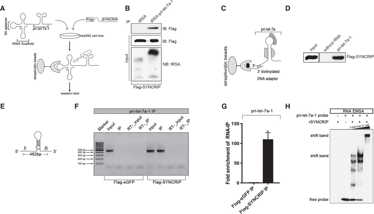
SYNCRIP binds to the pri-let-7a-1 both in vivo and in vitro. (A) A schematic representation of in vivo tRSA-RNA affinity assay using pri-let-7a-1 in fusion with tRSA tag in HEK293 cells overexpressed with FLAG-tagged SYNCRIP. (B) Detection of the presence of FLAG-SYNCRIP in the bound fraction of either tRSA (control) or tRSA-pri-let-7a-1 by in vivo tRSA-affinity assay. The eluate fraction and 5% of protein input were analyzed by immunoblot against FLAG tag. Five percent of RNA input was analyzed by northern blotting using DNA probe against tRSA tag. (C) A schematic representation of in vitro RNA affinity assay. Pri-let-7a-1 with short 5′ extension was in vitro transcribed and immobilized with streptavidin-coated Sepharose beads via 3′-biotinylated DNA adaptor, the sequence of which is reverse-complementary to the 5′ extension. (D) Pull-down of overexpressed FLAG-tagged SYNCRIP using pri-let-7a-1. The eluate fraction and 5% of protein input were analyzed by immunoblot against FLAG tag. 3′-biotinylated DNA adaptor immobilized on streptavidin-coated Sepharose beads was used as a control. (E) A schematic representation of primers used in RNA-IP PCR analysis. (F) RIP–PCR analysis of pri-let-7a-1 performed with anti-FLAG beads in HEK293 cells transfected with either flag-eGFP (negative control) or flag-SYNCRIP. (RT) No reverse-transcribed PCR. (G) qPCR analysis of pri-let-7a-1 bound with FLAG-SYNCRIP in RIP assay in HEK293 cells. The RIP–qPCR results were shown as fold enrichment compared with input. (H) RNA-EMSA assay using biotin-labeled pri-let-7a-1 (0.1 µM). Increasing concentration of purified recombinant his-SYNCRIP was indicated (0.1–0.4 µM).




