


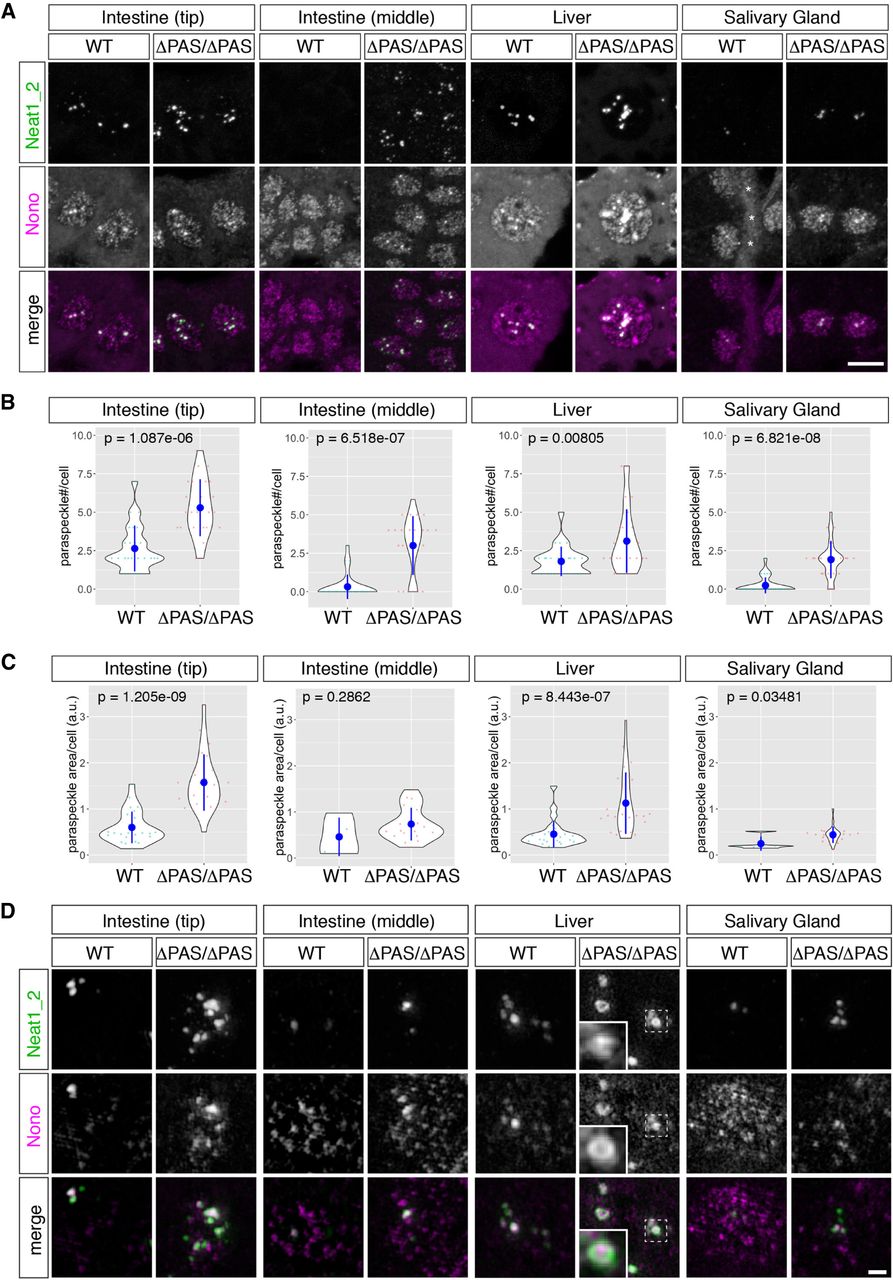
Simultaneous detection of Neat1_2 and the paraspeckle marker Nono in Neat1ΔPAS/ΔPAS mice. (A) Expression of Neat1_2 in the tip cells of the intestinal epithelium (top), the middle regions of the intestinal epithelium (middle), the liver, and the salivary gland, as revealed by probes that specifically detected Neat1_2 and by the simultaneous detection of the paraspeckle marker Nono. Note the marked increase in paraspeckle formation, which was identified by the accumulation of Nono, in the tip cells of the intestine epithelium and the liver cells in Neat1ΔPAS/ΔPAS (ΔPAS/ΔPAS) mice. Asterisks in the WT salivary gland indicate autofluorescent signals derived from the basement membrane. (B,C) Quantification of the number (B) and the areas (C) of paraspeckles per cell in the tip cells in the intestinal epithelium, the middle of the intestinal epithelium, the liver, and granular duct cells in the salivary gland. Note that the majority of the granular duct cells did not form paraspeckles in the WT mice. Twenty-five cells were randomly selected for the measurement from each tissue derived from two individual animals for each genotype. P-values were calculated by Wilcoxon's rank sum tests. Blue dots and bars represent the mean value and the standard deviation, respectively. (D) Simultaneous detection of Neat1_2 and Nono at high magnification using structural illumination microscopy. Dotted boxes indicate the area shown in the insets at higher magnification. Scale bars, 10 µm in A and B and 1 µm in D.




