


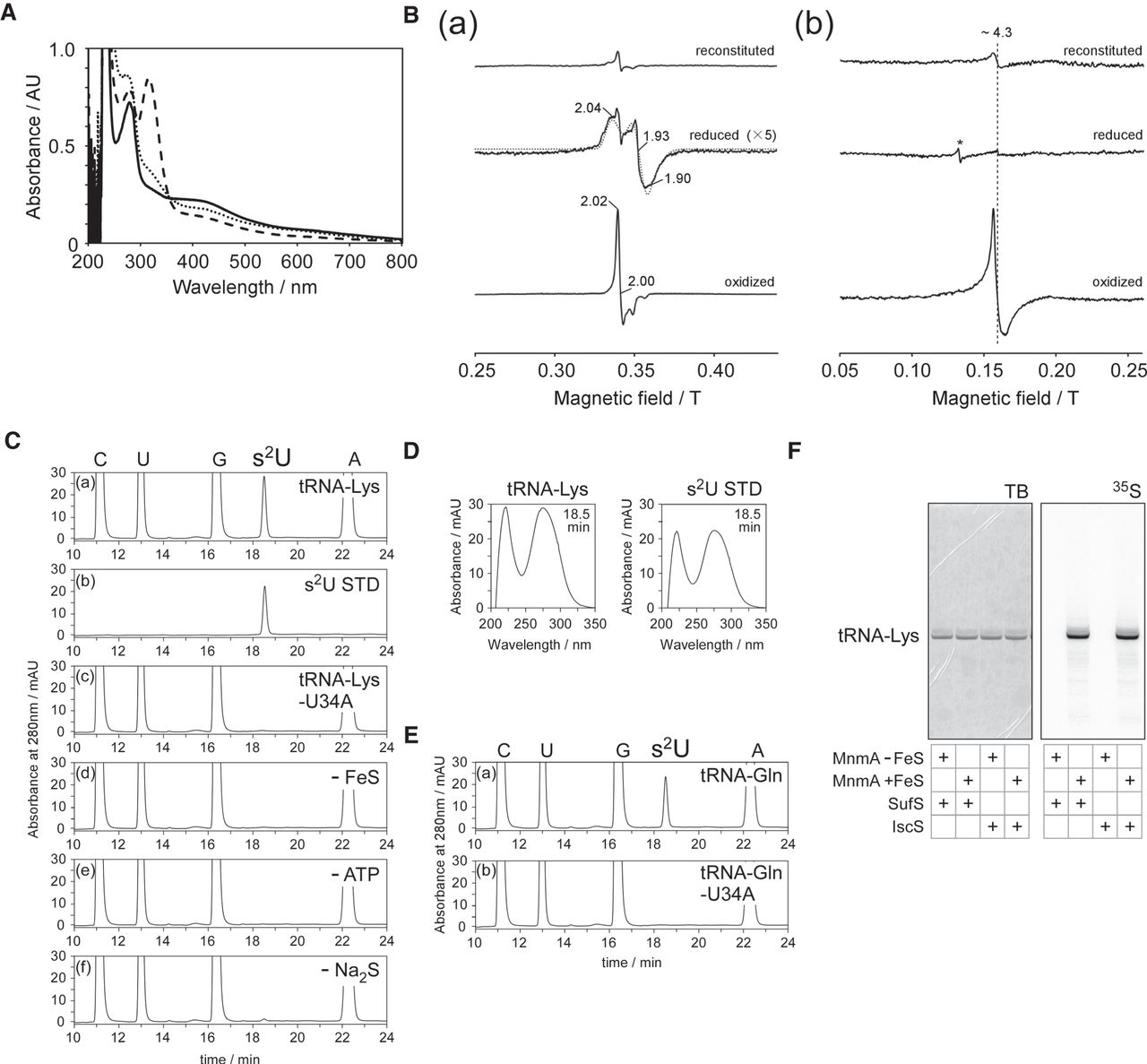
Characterization of T. thermophilus MnmA. (A) UV–VIS spectra of reconstituted (solid line), reduced (dashed line), and oxidized MnmA (dotted line). The samples of reduced and oxidized MnmA were prepared by adding 10 eq of sodium dithionite and ferricyanide solution, respectively, and diluted to 0.7 mg/mL in A buffer. The peak at around 310 nm in the reduced sample was derived from excess dithionite. (B) EPR spectra of reconstituted, reduced, and oxidized MnmA (∼0.7 mM) at 12 K in high (a) and low (b) magnetic fields. The principle g-values are shown in the figure. All spectra acquired under a high magnetic field were subjected to subtraction of the spectrum acquired using buffer alone. A simulated spectrum of the [4Fe–4S]1+ cluster is also presented in (a), represented by a dotted line. The asterisk in (b) shows an unknown signal with g ∼ 5.7. Detailed conditions: microwave frequency = ∼ 9.59 GHz, microwave power = 1 mW, 100 kHz modulation amplitude = 10 G, time constants = 41 ms, number of scans = 4, and temperature = 12 K. (C) Nucleoside analysis of transcribed tRNALys from in vitro reactions with holo-TtMnmA. The modified nucleosides of the reacted RNA were analyzed by HPLC. (a) tRNA (450 pmol) was reacted at 60°C for 20 min with 19 pmol of MnmA in the presence of 0.1 mM sodium sulfide, 2.5 mM ATP, and 0.1 mM DTT. (b) s2U standard. (c) tRNALys with a U34A mutation was used. (d) Apo-MnmA was used. In (e) and (f), ATP and sodium sulfide were omitted, respectively. (D) UV spectra of s2U detected at 18.5 min in tRNALys (C[a]) and authentic s2U (C[b]). (E) Nucleoside analysis of transcribed tRNAGln from in vitro reactions. Wild-type (a) and U34A mutant (b) molecules were analyzed. The assay conditions were the same as in C. (F) [35S]-Cys-derived 35S-sulfur incorporation into s2U. tRNALys (450 pmol) was reacted at 60°C for 30 min with 19 pmol of apo- or holo-MnmA and 75 pmol of IscS or SufS in the presence of 20 µM [35S]-Cys, 20 µM pyridoxal phosphate, 2.5 mM ATP, and 0.1 mM DTT. The reacted RNA was separated by 10% denaturing PAGE and then stained with toluidine blue (TB; left), after which the 35S radioactivity was visualized (right).




