


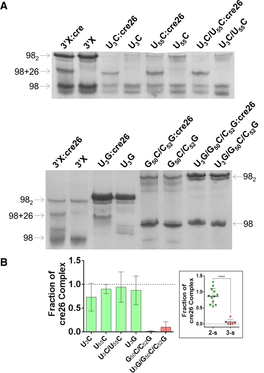
Gel electrophoresis analysis of 3′X domain molecules and their complexes with the 5BSL3.2 hairpin cre26. (A) Native gels comparing the electrophoretic mobility of wild-type and mutant U3C, U55C, U3C/U55C, U3G, G50C/C52G, and U3G/G50C/C52G 3′X domain molecules in the absence and presence of one molar equivalent of cre26. The arrows indicate the position of the monomer (98), homodimer (982), and heterodimer (98 + 26) species. (B) Quantification of the interaction between mutant 3′X domain molecules and cre26, obtained by measuring the fraction of 3′X:cre26 complexes (98 + 26) relative to total 3′X RNA. The results were normalized with respect to the wild-type sequence, which was assigned a value of 1, and the bars represent the average and standard deviation of three independent experiments. Mutants experimentally verified to adopt the wild-type two-stem conformation are represented in green, whereas mutants adopting a different structure are indicated in red. In the scatter plot shown in the inset, the individual values of the two-stem and three-stem mutants are indicated, together with means and standard deviations. The differences between two-stem and three-stem mutants are significant in all cases (P < 0.005). Conditions: 10–40 µM RNA, TBM running buffer (2 mM MgCl2).




