


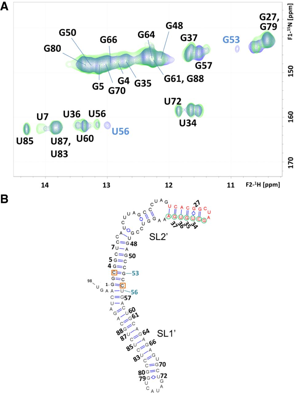
NMR spectroscopy analysis of the U3C/U55C 3′X mutant. (A) 1H–15N HSQC spectrum of U3C/U55C at low ionic strength (green), superposed with that of the wild-type 3′X domain (blue) acquired under the same temperature and ionic conditions. All assignments appear in black except for those of wild-type G53 and U56 HN crosspeaks, which do not overlap with U3C/U55C mutant signals and are labeled in blue. In the wild-type domain, the U56 signal is doubled due to conformational exchange. (B) Secondary structure model of the U3C/U55C mutant domain supported by the NMR data. DLS nt are depicted in red, k motif nt are indicated with green circles, and the U3C and U55C mutations stabilizing the lower stem of subdomain SL2′ are marked with red squares. Residues with assigned HN resonances are numbered. Conditions: 64 µM U3C/U55C, 0 mM NaCl/MgCl2, 27°C.




