


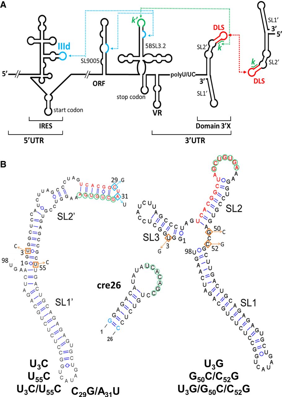
Secondary structure of HCV 3′X wild-type and mutant domains analyzed in this study. (A) Schematic representation of the secondary structure of the 5′ and 3′ regions of the HCV RNA genome. Functional RNA–RNA interactions are indicated by blue and green dotted arrows. The red arrow represents an intermolecular contact between palindromic DLS sequences that promotes viral RNA homodimerization. (B) Predicted secondary structure of the 98-nt-long 3′X domain and the 26-nt-long apical portion of the 5BSL3.2 domain (cre26). The U3C, U55C, and U3C/U55C 3′X domain mutants stabilize the two-stem conformation observed for the wild-type sequence, whereas the U3G, G50C/C52G, and U3G/G50C/C52G mutants were designed to favor an alternative domain structure formed by three stems. The C29G/A31U mutant, previously shown to adopt the two-stem conformation (Cantero-Camacho and Gallego 2018), contains a double mutation that disrupts the DLS palindrome and blocks 3′X domain dimerization. The blue-colored nt in cre26 indicates changes relative to the wild-type sequence introduced to increase transcription yield. The k sequence motif within domain 3′X (indicated with green circles) has been shown to establish a distal interaction involving Watson–Crick pairs with a complementary k′ sequence (green circles) in the apical loop of 5BSL3.2. The 16-nt palindromic DLS motif (depicted with red nt) includes the k tract and is universally conserved among all HCV isolates. The two- and three-stem conformations of the 3′X domain expose differently the k and DLS motifs.




