The origin of the high stability of 3′-terminal uridine tetrads: contributions of hydrogen bonding, stacking interactions, and steric factors evaluated using modified oligonucleotide analogs

(Downloading may take up to 30 seconds. If the slide opens in your browser, select File -> Save As to save it.)

Click on image to view larger version.

FIGURE 3.
FIGURE 3.

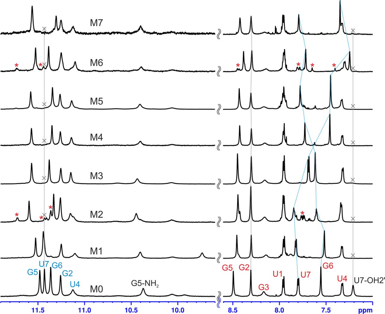
The 1H-NMR spectra (imino and aromatic regions) of G-quadruplexes M0–M7 recorded in Na+ containing conditions at 25°C. The blue dotted lines follow some among the resonances with the greatest chemical shift changes. Additional NMR signals from minor species present in some of the samples are marked with asterisks.

This Article

  1. RNA 26: 2000-2016

