


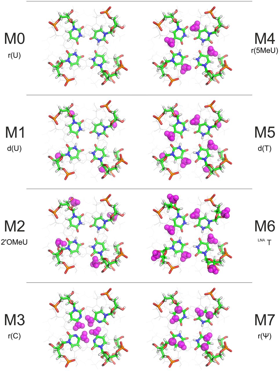
FIGURE 2.
The chemical modifications of the 3′-terminal uridine residue (G-quadruplexes M0–M7). The purple spheres indicate the sites at which groups of atoms were added, removed, or substituted depending on the modification. The figure depicts each modified G-quadruplex in an idealized conformation with a well-formed “reversed” U-tetrad, which many among them do not assume in reality (see text).




