


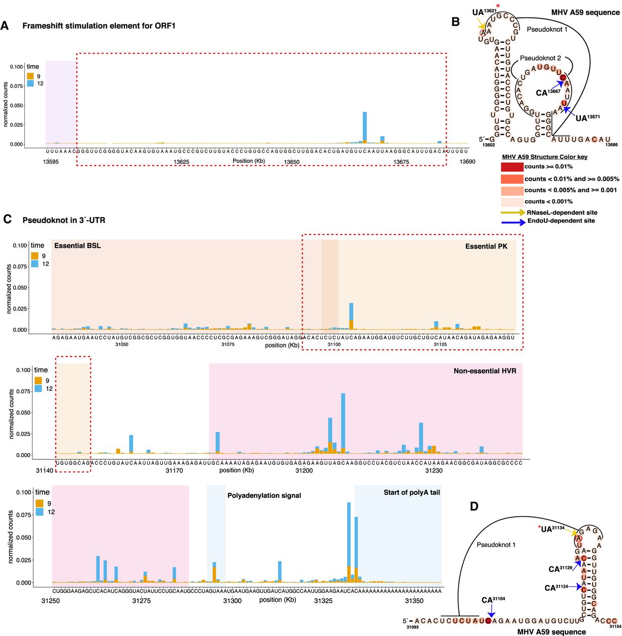
MHV secondary structures associated with RNase L–dependent and EndoU-dependent cleavage sites. (A,C) Nucleotide resolution graphs displaying normalized counts by position for the regions encompassing secondary structure predictions. (B,D) Secondary structures of frameshift stimulation element (B) and MHV 3′-UTR pseudoknot (D), generated using available consensus alignment and the R-scape program (Rivas et al. 2017). MHV A59 sequence mapped to consensus secondary structures using available covariation model and the Infernal program (Nawrocki and Eddy 2013). Base coloring of MHV A59 sequence based on normalized cDNA reads as indicated in key for 12 hpi in wt BMM infected with MHV(V). *Base RNase L–dependent cleavage activity is increased in PDEmut or EndoUmut infection as compared to MHV(V) infection.




