


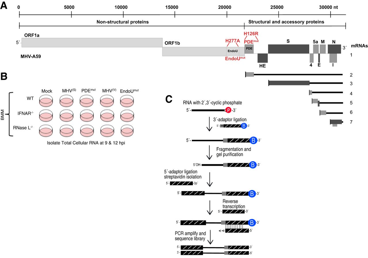
Coronavirus RNA genome and experimental approach. (A) MHV RNA genome highlighting two mutations: His to Arg mutation in the MHV phosphodiesterase domain active site (PDEH126R), and a His to Ala mutation in the MHV EndoU domain active site (EndoUH277A) (Roth-Cross et al. 2009; Kindler et al. 2017). MHV proteins are categorized as nonstructural (light gray), accessory (dark gray), and structural (black). Subgenomic mRNAs 2–7, produced during infection, are illustrated. (B) Bone marrow–derived macrophage (BMM) from wt, IFNAR−/−, and RNase L−/− mice were mock-infected or infected with wt MHV (MHV(S) and MHV(V)), the PDEmut, or EndoUmut for 9 and 12 h (Zhao et al. 2012; Kindler et al. 2017), after which total cellular RNA was isolated for cyclic phosphate sequencing. (C) Schematic of cyclic phosphate sequencing; protocol adapted from Schutz et al. (2010).




