


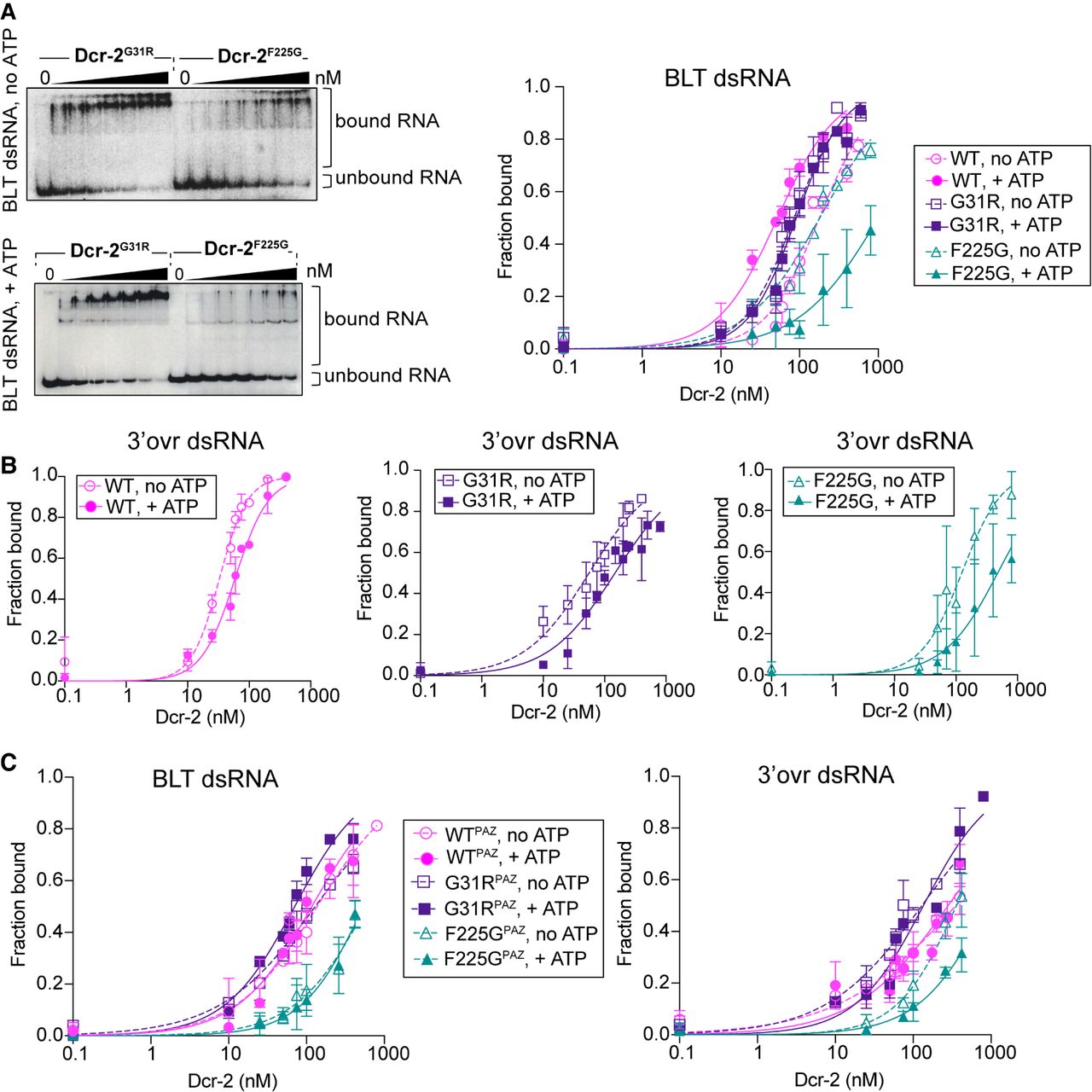
Effects of helicase mutations on dsRNA binding. (A) 32P-labeled BLT 52-dsRNA was incubated with Dcr-2WT, Dcr-2G31R, or Dcr-2F225G, ±5 mM ATP as indicated on left of gel. EMSA data were quantified and fit using the Hill formalism (right panel, error bars, SD, n = 3–5), with fraction bound = 1/(1 + (Kdn/[P]n)), where Kd = binding affinity and [P] = protein concentration. (B) Quantification of EMSAs (see Supplemental Fig. S5) for 3′ovr dsRNA binding to Dcr-2WT, Dcr-2G31R, or Dcr-2F225G ±5 mM ATP. (C) Quantification of EMSAs (see Supplemental Fig. S5) for BLT (left) and 3′ovr (right) dsRNA binding with Dcr-2WT, Dcr-2G31R, or Dcr-2F225G, with two mutations in the PAZ domain (R943A, R956A) ±5 mM ATP. Data were quantified (error bars, SD; n = 3–5) and plotted as in A. Dcr-2F225G,PAZ was only purified through Strep-Tactin affinity chromatography. All Dicer proteins contained a wild-type RNase III domain, and some assays showed cleavage products (see Supplemental Fig. S5). Control experiments using proteins with mutations in the RNase III domains showed no significant differences in calculated Kd values.




