


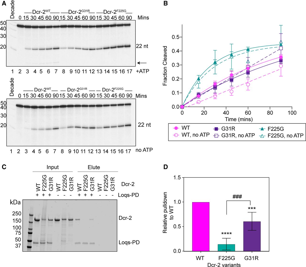
Effects of helicase mutations on 3′ovr dsRNA cleavage and Loqs-PD binding in vitro. (A) PhosphorImages show representative 16% denaturing polyacrylamide gels monitoring cleavage of 3′ovr dsRNA by Dcr-2 with 5mM ATP (top), and without ATP (bottom). The arrow points to 15 nt product. (B) Quantification of data as in A (mean ± SD, n = 3), fit to a pseudo first-order equation, y = y0 + A (1 − e−kt), where A = amplitude of rate curve, y0 = baseline (∼0), k = kobs (first-order rate constant), and t = time (min). (C) Dcr-2 variants, as indicated, were incubated for 60 min ± His-tagged Loqs-PD. Representative 4%–15% SDS-polyacrylamide gel stained with Coomassie Brilliant Blue shows input proteins and proteins eluted after pulldown. Far left lane shows molecular weight markers with sizes labeled to the left of the gel. (D) Graph shows quantification of pulldown assay relative to WT protein. n = 4 (three different protein preparations); error bars, SEM; unpaired Student's t-test as compared to Dcr-2WT: (***) P < 0.001; (****) P < 0.0001. Additional comparison between Dcr-2G31R and Dcr-2F225G: (###) P < 0.001. Dcr-2G31R and Dcr-2F225G purified through Strep-Tactin affinity chromatography only.




