


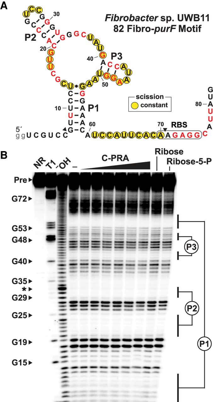
In-line probing analysis of a Fibro-purF motif RNA is consistent with the predominant secondary structure features. (A) Sequence and secondary structure of the Fibro-purF motif RNA from Fibrobacter sp. UBW11. Lowercase letters identify nucleotides added to increase in vitro transcription efficiency. Red nucleotides are conserved >97% as noted in the consensus sequence (Fig. 2A). Yellow circles identify nucleotide positions that undergo spontaneous RNA cleavage regardless of ligand addition as revealed by an in-line probing assay depicted in B. Arrowheads identify the locations where the mapping of in-line probing data from B began and ended. (B) Denaturing (8 M urea) polyacrylamide gel electrophoresis (PAGE) analysis of in-line probing reactions conducted with 5′ 32P-labeled 82 Fibro-purF RNA in the absence (−) or presence of C-PRA (tested at log increments from 10 nM to 1 mM), ribose (1 mM), or ribose 5-phosphate (1 mM). NR, T1, and −OH indicate no reaction, partial digestion with T1 ribonuclease (cleaves after every G), and partial digestion under alkaline conditions (cleaves after every nucleotide), respectively. Bands corresponding to precursor 82 Fibro-purF RNA (Pre) and RNase T1 digestion products are annotated. The asterisk denotes an electrophoretic mobility compression that distorts the location of bands in the 31- to 33-nt size range.




