


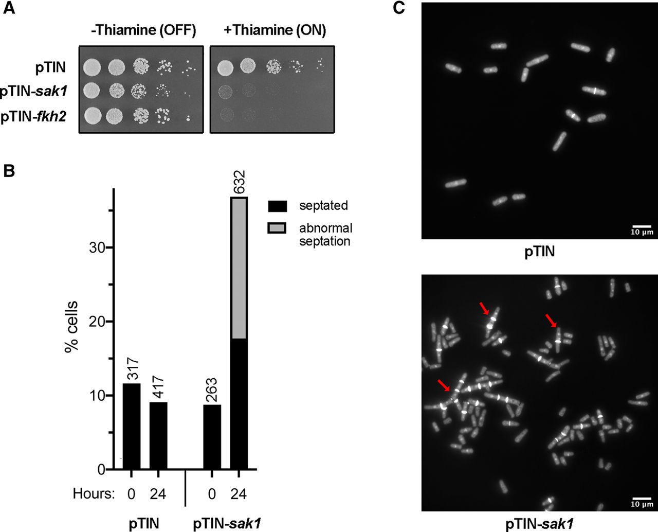
Overexpression of the mitotic transcription factors Sak1 and Fkh2. (A) Single transformants of WT cells bearing the pTIN, pTIN-sak1, or pTIN-fkh2 plasmids were propagated in Leu− PMG medium without thiamine. Serial dilutions of cells were spotted on Leu− PMG medium with or without 15 µM thiamine and were incubated at 30°C for 3 or 4 d, respectively. (B) A representative culture of WT cells bearing either the pTIN empty vector or pTIN-sak1 plasmid were grown at 30°C in Leu− PMG medium lacking thiamine. An aliquot of the cultures was fixed (time 0) and the remaining volume of cultures was adjusted to 15 µM thiamine. The cultures were incubated at 30°C and an aliquot of each culture was fixed after 24 h. Fixed samples were processed for DAPI and Calcofluor White staining and the proportion of septated cells was quantified by fluorescence microscopy. The total number of cells counted for each sample is indicated above the bar. The Z-test two tailed P-values comparing the proportion of total septated cells of pTIN and pTIN-sak1 samples are 0 h: 0.30; 24 h: 9.18 × 10⁻²⁴. (C) Fluorescence images (DAPI and Calcofluor) of cells bearing the indicated plasmids (described in Fig. 5B) under de-repressed (+thiamine) conditions at 24 h. Red arrows indicate examples of cells with aberrant mitosis.




