


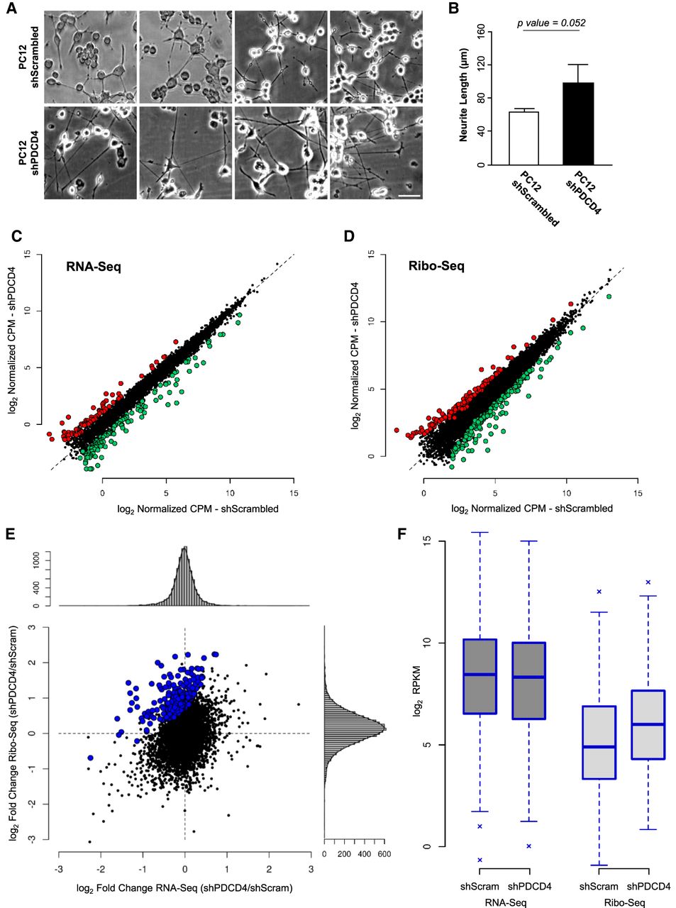
Translatome and steady-state transcriptome levels of expression for mRNAs was quantified genome-wide by Ribo-seq and RNA-seq, respectively, in absence (shPDCD4) and presence (shScrambled) of PDCD4 achieved by lentiviral transfection in neuron-like differentiated PC12 cells. (A) To explore if PDCD4 modulates neurite outgrowth also in differentiated PC12 cells, we compared neurite length in differentiated PC12 growing in presence and absence of PDCD4. Three independent cultures were contrasted and illustrative fields are shown (scale bar, 50 µm). (B) Quantification of neurite length of A is shown and an increase of almost 1.6× in absence of PDCD4 is detected, with a marginal trend toward significance (P = 0.052, Student's t-test, n = 3 independent cell cultures with an average of 80 neurites considered by replicate, error bars: SD). (C) Scatter plot showing PDCD4 regulation at the level of transcriptome evaluated by RNA-seq. Red and green dots indicate differentially expressed genes, up- and down-regulated, respectively (|fold change| > 2 and P < 0.05 estimated by edgeR). (D) Same as C but for PDCD4 regulation at the level of translatome evaluated by Ribo-seq. (E) Fold changes (shPDCD4/shScrambled) at transcriptome and translatome levels are contrasted for detected genes. Possible PDCD4 translational targets are those that present a significant increase in translational efficiency (P < 0.05 estimated by Xtail), and are indicated in blue (267 mRNAs). Along the scatter plot, vertical and horizontal histograms show fold change values distribution estimated by Ribo-seq and RNA-seq. (F) RPKM gene expression levels for PDCD4 translational targets indicated in blue in E are shown for the two compartments (RNA-seq and Ribo-seq) in each condition.




