


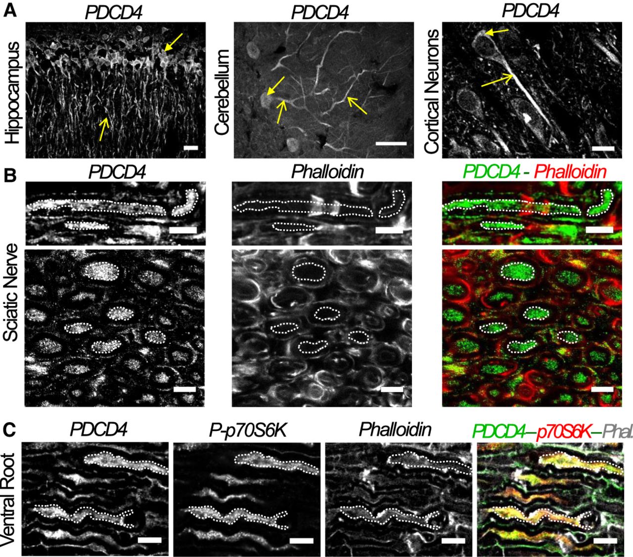
PDCD4 is expressed in dendrites and axons of the central and peripheral nervous system. (A) Immunohistochemistry assays show the distribution of PDCD4 protein at different types of neurons including CA1 hippocampal neurons, Purkinje of cerebellum and cortical neurons of prefrontal cortex of adult rats. Cell bodies are indicated by filled arrows and axons (or dendrite for Purkinje neurons) by unfilled arrows (scale bar, 20 µm). (B) PDCD4 protein is also detected in peripheral axons, like sciatic nerves, by immunohistochemistry. The images above correspond to longitudinal sections and the images below to transversal sections. Examples of axonal regions are highlighted in white dotted ROIs (scale bar, 10 µm). (C) Longitudinal sections of ventral roots. Examples of axonal regions are highlighted with white dotted ROIs. A partial colocalization between PDCD4 and p70-S6K signals is detected (scale bar, 5 µm).




