


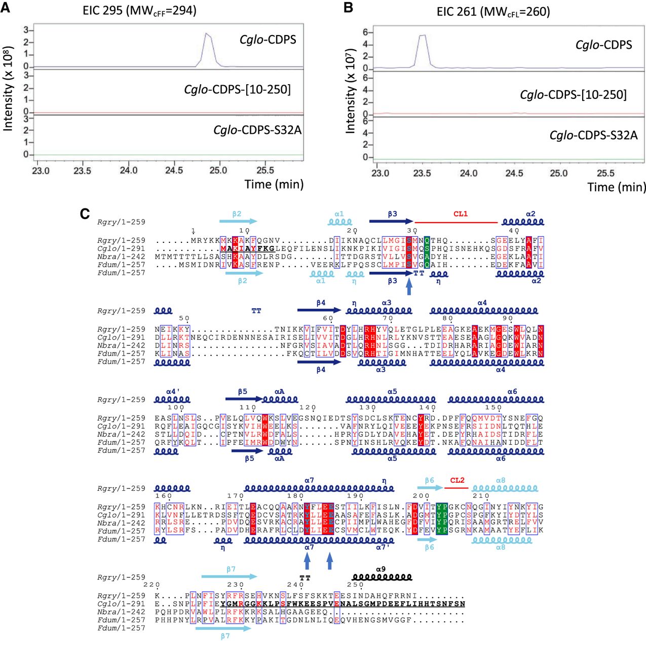
Cglo-CDPS. (A,B) Determination of the cyclodipeptide-synthesizing activities of Cglo-CDPS by LC-MS analyses. Extracted ion chromatograms (EIC) corresponding to the m/z of cFF (A) and cFL (B) from the supernatants of E. coli cells expressing wild-type Cglo-CDPS (blue) and its variants Cglo-CDPS-[10-250] (red) and Cglo-CDPS-S32A (green) are displayed. (C) Sequence alignment of Cglo-CDPS with three other XYP-CDPSs of known 3D structure. The figure was drawn with Espript (Gouet et al. 1999). Secondary structures of Rgry-CDPS are indicated at the top of the alignment and those of Fdum-CDPS are indicated at the bottom. The first half of the Rossmann fold is colored in cyan and the second half of the Rossmann fold is colored in blue. Residue characteristics of the XYP-subfamily are in green boxes. Other important residues (S29, E185, Y181, Rgry- numbering) are shown in bold blue and indicated by a vertical arrow. Residues displayed in bold in β2 and β7 of Cglo-CDPS sequence indicate the deleted region in the Cglo-CDPS-[10-250] variant.




