


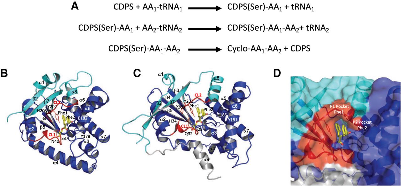
Catalytic cycle and binding of the dipeptide to CDPS. (A) Catalytic cycle of CDPS. The aminoacyl-enzyme intermediate is bound to a serine residue. (B) Structure of Snou-CDPS (formerly called AlbC) bound to a Phe–Phe dipeptide. The model is deduced from the structure of Snou-CDPS bound to a reaction intermediate (PDB ID code 4Q24) (Moutiez et al. 2014a; Schmitt et al. 2018). The first part of the Rossmann fold is colored in cyan and the second part is in dark blue. The two catalytic loops CL1 and CL2 are in red. Residues important for catalysis are shown in sticks. The dipeptide bound to the catalytic serine is in yellow stick. Snou-CDPS is a member of the NYH-CDPS subfamily (Jacques et al. 2015; Bourgeois et al. 2018). (C) Structure of Rgry-CDPS bound to a Phe–Phe dipeptide. The same color code as in view B is used. Rgry-CDPS is a member of the XYP-CDPS subfamily. (D) Closeup of the Rgry-CDPS dipeptide binding site from view C. The molecular surface is represented. The view shows the two substrate binding pockets named P1 and P2 (see text).




