


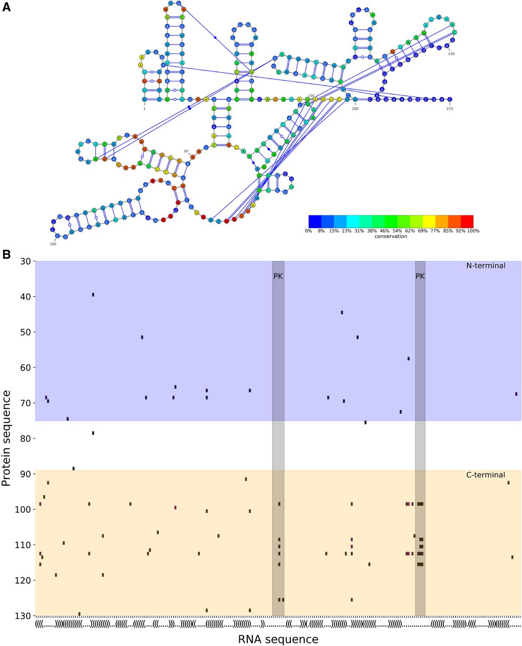
FIGURE 1.
(A) GII secondary structure and its sequence conservation. The last 25 positions have no conservation levels because they are excluded from the alignment (see section “Group I intron” in Materials and Methods). (B) Positions of significant DCA scores (≥4σ above average) between the StpA protein (y-axis) and the GII RNA (x-axis). The RNA axis is labeled with the secondary structure in parentheses notation. The blue region is the amino terminal of StpA and the orange region its carboxyl terminal. The pale gray regions are the pseudoknot (PK) of GII. The last 25 positions of GII are omitted (see section “Group I intron” in Materials and Methods).




