


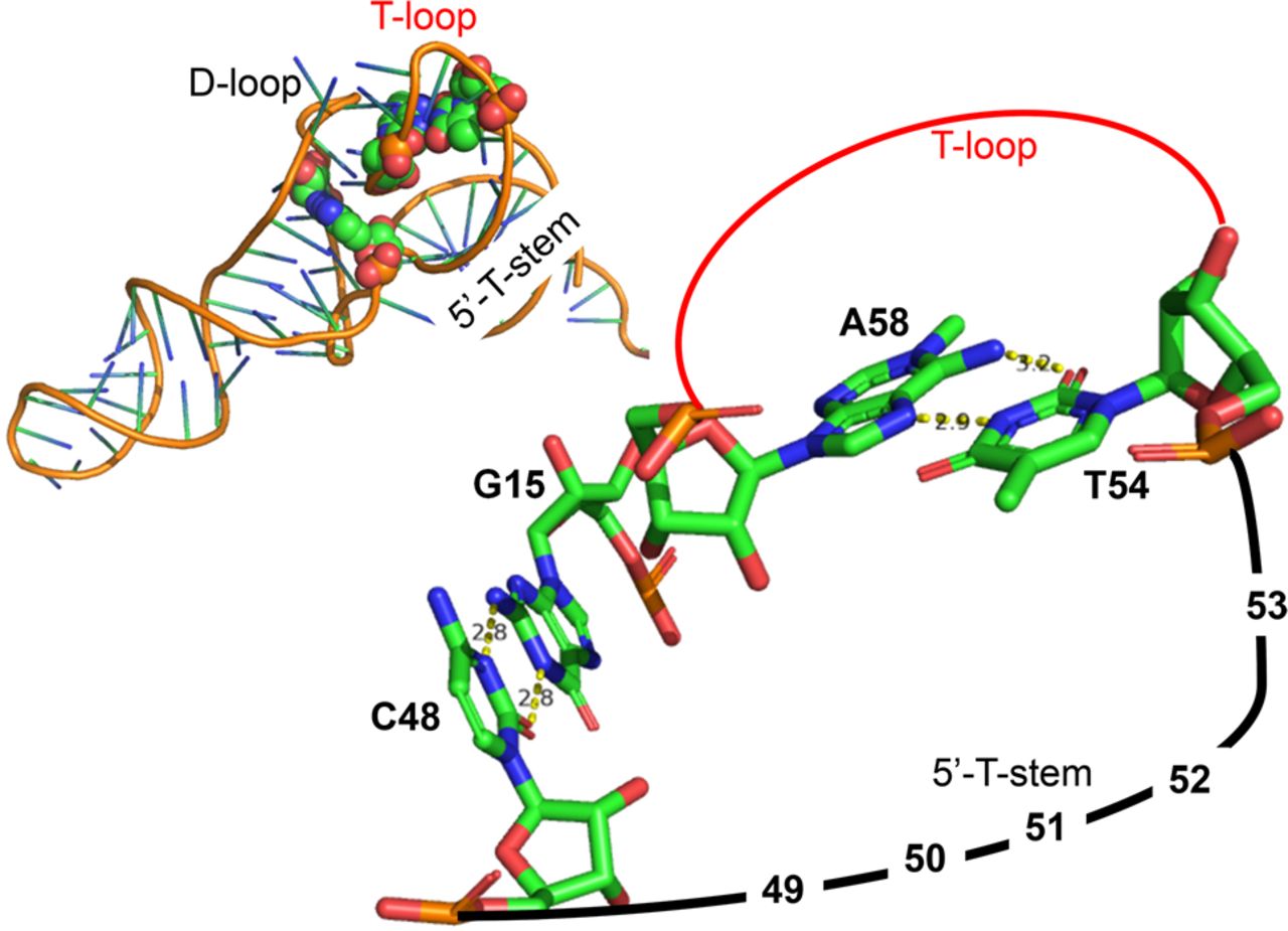
FIGURE 4.
On the top left, a tRNA structure (PDB 1EHZ from Shi and Moore 2000) is shown with the two non-Watson–Crick pairs 15/48 (linking the beginning of the D-loop to the end of the variable loop) and 54/58 (closing the T-loop) highlighted with van der Waals spheres. On the right, a simplified diagram shows that both 48 and 54 are on the same strand so that a change from a pyrimidine to a purine would draw toward the reader the strand (with probably accompanying movement of the T-helix). Drawings made using PyMOL (PyMOL 1.7.7.6—Incentive Product © Schrodinger LLC).




