


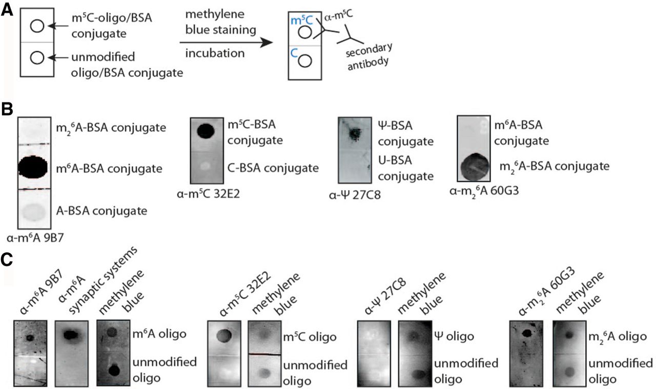
Antibody specificity against base modifications using dot blot assays. (A) Scheme of the dot blot experimental setup. Unmodified/modified BSA conjugates or oligonucleotides (m5C or C as examples) were spotted on a nylon membrane. After cross-linking and blocking, the membrane was incubated with the primary and secondary antibodies. Methylene blue stains nucleic acids and was used as a loading control. (B) Dot blot experiments, for which modified and unmodified BSA-nucleoside conjugate was used to assess the specificity of the antibodies α-m6A 9B7, α-m5C 32E2, α-Ψ 27C8, and α-m26A 60G3. For m6A, also the m26A-BSA conjugate was spotted to investigate specificity between the two very similar modifications. (C) Dot blot experiments using a modified and a nonmodified 12-mer RNA oligonucleotide as negative control. The antibody staining is shown on the left and the methylene blue staining as a loading control on the right. The dot blots for the antibodies α-m6A 9B7 and Synaptic Systems, α-m5C 32E2, α-Ψ 27C8, and α-m26A 60G3, are depicted.




