


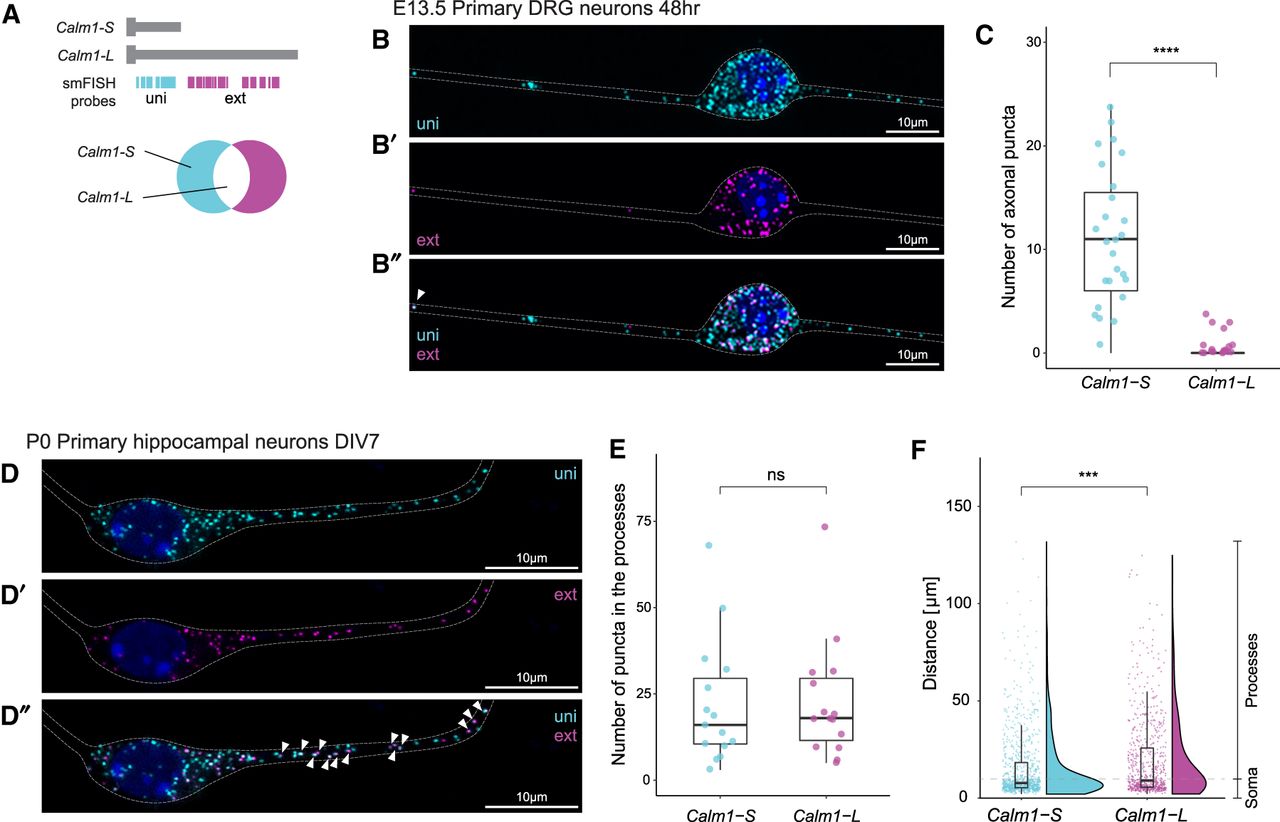
Subcellular localization of Calm1-L. (A) Diagram showing RNAscope smFISH probe locations. When uni and ext images are merged, Calm1-L isoforms are shown as colocalized (white) puncta. (B) smFISH showed robust axonal localization of Calm1-S (uni), but Calm1-L (ext/magenta in B′ or white punta in B″) was observed in DRG axons. (C) When the number of axonal uni or ext puncta was counted, uni signals were robustly found in the axons of DRG neurons but little to no ext signals were found in the same regions. Significance was determined using a t-test, (****) P < =0.0001. n = 30 neurons. (D) In primary hippocampal neurons, both Calm1-S and Calm1-L were observed in soma and neuronal processes. (E) The number of puncta corresponding to Calm1-S and Calm1-L transcript in the hippocampal processes was not found to be significantly different. Significance was determined using a t-test, ns: P > 0.05. n = 15 neurons. (F) Analysis of the distance of travel for all the Calm1-S and Calm1-L signals showed the overall distribution is different between two mRNA isoforms. Analysis done in n = 15 neurons (Calm1-L 684 puncta, Calm1-S 907 puncta). Significance was determined using a Wilcoxon test, (***) P < =0.001.




