


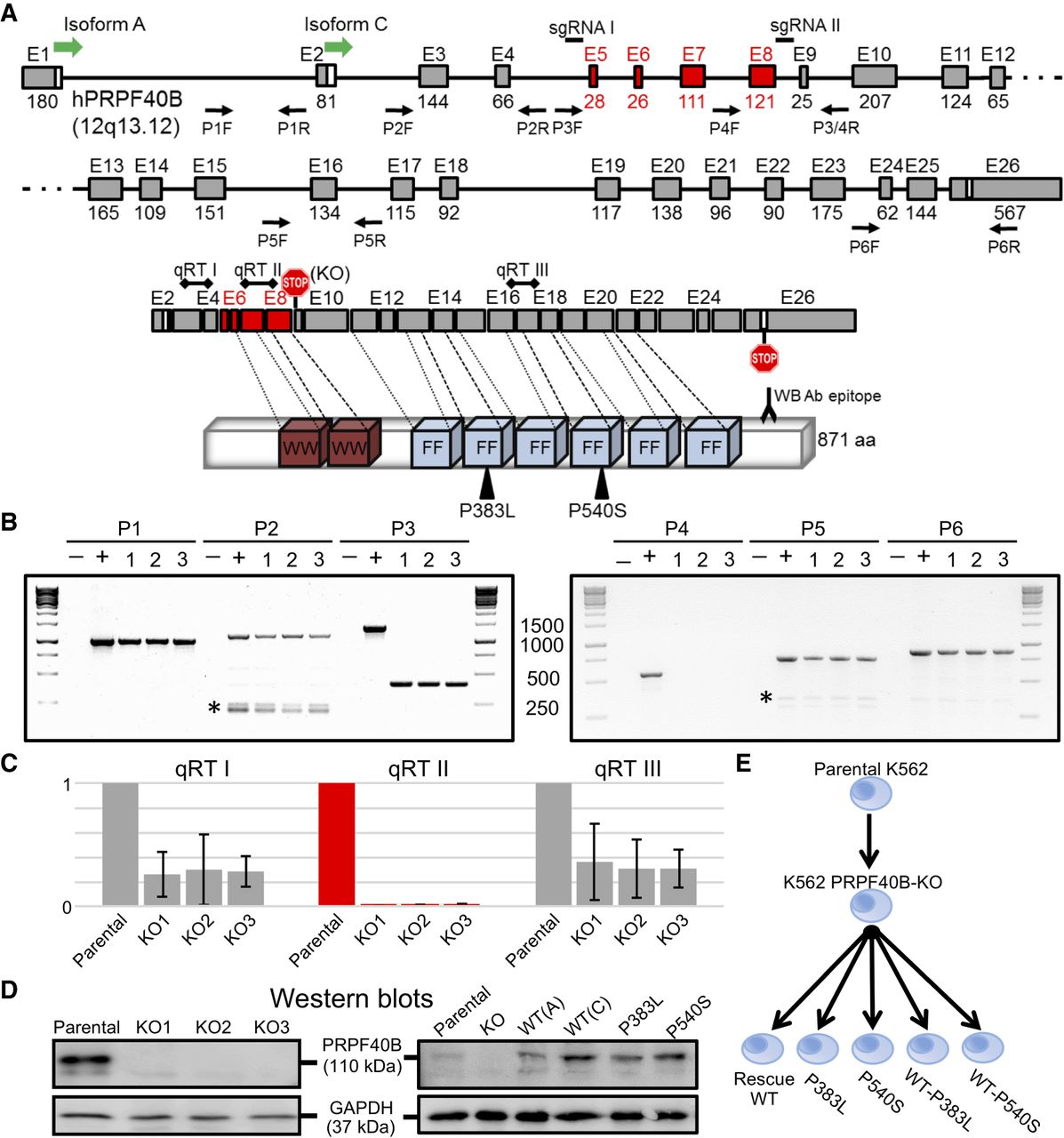
Design and validation of PRPF40B-KO and rescue in K562 cells. (A) Schematic of PRPF40B gene, mRNA, and protein. We depict the PRPF40B gene with exons on scale, with the translation start sites of isoforms A and C. We indicate the position of the sgRNAs for the CRISPR/Cas9 KO that flank the deletion including exons 5–8 in red, and the pairs of primers for genomic PCR validations. The mRNA shows the natural stop codon in last exon 26, the termination codon in exon 9 introduced by the deletion (KO), and the positions of the three amplimers for the real-time RT-PCR. mRNA exons are aligned to their WW and FF protein domains, with the MDS mutations and the monoclonal antibody epitope for western blot. (B) PCR genotyping of PRPF40B-KO clones 1–3. The PCR number matches the primer pairs in Figure 1A. (–) Negative control, water template; (+) positive control, parental K562 genomic DNA. Ladders are shown on each side of gel, with the band sizes in base pairs in the middle. PCRs 1, 2, 5, and 6 fall outside the deletion fragment and are detected in all KO clones. PCR 3, with primers flanking the deletion, shows the full-length band in parental cells, and a much smaller band in the KO clones because of the deletion. PCR 4, with one primer inside the deletion, only generates a band in parental but not KO cells. The identity of specific bands was confirmed by sequencing. Asterisk, unspecific amplimers. (C) Real-time RT-PCR measurement of PRPF40B RNA levels for the qRT I–III regions in Figure 1A. We plotted PRPF40B expression ratios relative to β-actin and with parental K562 values normalized to 1. (D) Representative western blots of the parental K562 and three PRPF40B-KO clones (left), and of overexpression samples with WT (isoforms A or C) and mutant protein (right), both with GAPDH as loading control. (E) Diagram of the samples with altered PRPF40B expression, including KO by CRISPR/Cas9 in K562 cell line, and rescue by overexpression in the KO cells of PRPF40B either as WT (Rescue WT) or the indicated MDS mutations alone or mixed with WT at 1:1 ratio. We performed RNA-sequencing with all these seven conditions.




