


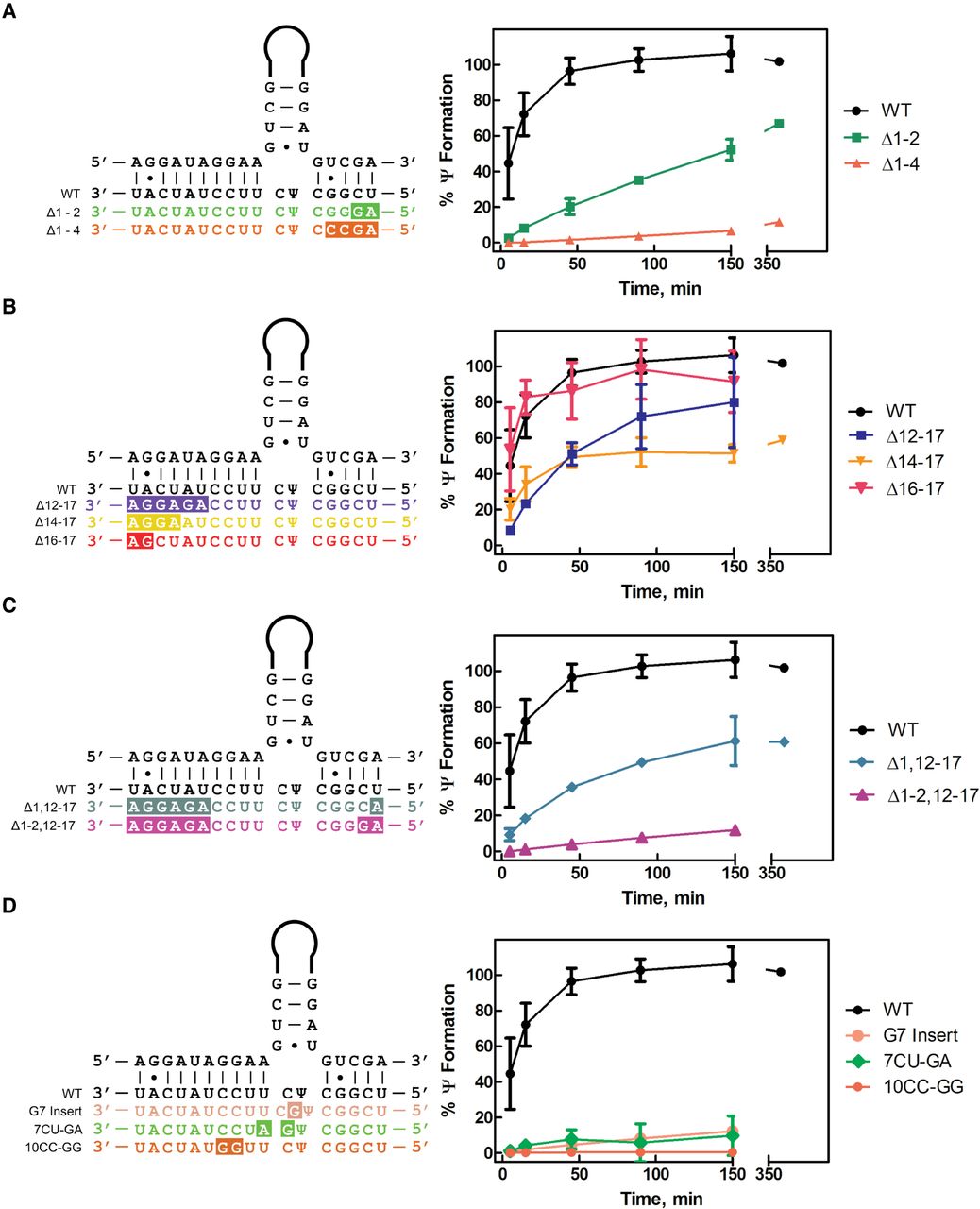
In vitro pseudouridylation of 3′ hairpin short substrate variants by the snR34 H/ACA snoRNP. Multiple turnover pseudouridylation reactions were performed by mixing 500 nM of each [3H-C5] uridine-labeled 3′ substrate RNA with 50 nM reconstituted snR34 H/ACA snoRNP. Location and sequence of substitutions are highlighted in the substrate RNA sequences on the left side, and the results of the tritium release assays are shown on the right side. (A) Modification of substrate RNA variants with mismatches in the 3′ side of the pseudouridylation pocket. The wild-type substrate (black circles), Δ1–2 substrate (green squares), and Δ1–4 substrate (orange triangles) are compared. (B) Modification of substrate RNA variants with mismatches in the 5′ side of the pseudouridylation pocket. The wild-type substrate (black circles), Δ12–17 substrate (indigo squares), Δ14–17 substrate (yellow inverted triangles), and Δ16–17 substrate (red, bold inverted triangles) are compared. (C) Modification of substrate variants with mismatches in both sides of the pseudouridylation pocket. The wild-type substrate (black circles), Δ1,12–17 substrate (teal diamonds), and Δ1–2,12–17 substrate (magenta triangles) are compared. (D) Modification of substrates with mismatches at internal sites in the pseudouridylation pocket. The wild-type substrate (black circles), 10CC-GG substrate (orange circles), 7CU-GA substrate (green diamonds), and G7 insert substrate (peach circles) are compared. Mean and standard deviation of three replicates are shown.




